Granuloma annulare
Exam relevance: in 3 of more than 50 board exam reports · rank 45
- Specialty
- Dermatology · Inflammatory skin diseases
- Images
- Clinical 3 · Dermoscopy 1 · Histology 1
- In the app
- 7 flashcards
- Last updated
- 10/2026 · Dr. Pascal Bafteh
Contents
Images (5)
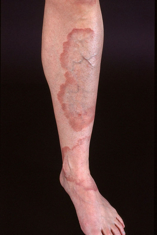
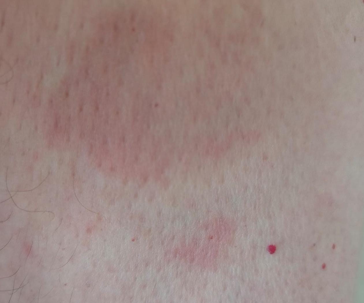
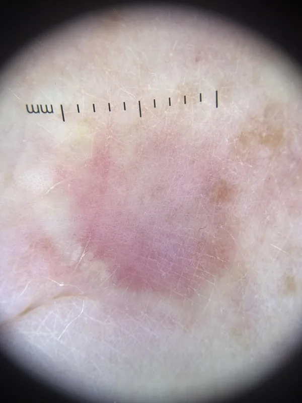 Dermoscopy
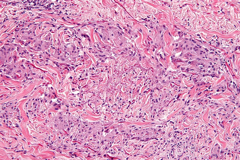 Histology
Definition
Definition & Classification
- Granuloma annulare (GA) = chronic-recurrent, granulomatous skin disease with annular plaques.
- Clinical Subtypes: localized + generalized + subcutaneous as well as perforating.
Aetiopathogenesis
Pathogenesis
- Immunological Reaction due to misdirected immune response to various triggers:
- Comorbidities: Diabetes mellitus, thyroid autoimmunity, hyperlipidemia (especially in generalized form), HIV.
- Cellular Infiltration: T-cells + macrophages + fibroblasts infiltrate the dermis and form granulomatous lesions.
- Cytokines / Chemokines: IL-2, IL-4, IL-10, IL-12, TNF-α, IFN-γ — Th1 / Th17 + Th2 mixed pattern with fibroblast activation.
- Fibroblast activation: Production of degraded collagen + mucin in the center of the granuloma, annular growth.
Clinical features
Clinical Presentation
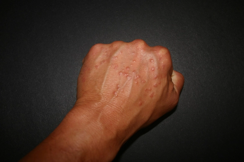
- Classic (localized): annular erythematous papules on dorsa of hands/feet + extensor surfaces.
- Generalized: > 10 lesions, often trunk + extremities, more common in adulthood, associated with diabetes mellitus and hyperlipidemia.
- Subcutaneous: painless subcutaneous nodules, often on the tibia / shin in children — DD to rheumatoid nodules important (Subcutaneous nodules of childhood).
- Perforating: umbilicated papules with crusts; rare.
- Patch / patchy: flat brownish-erythematous plaques.
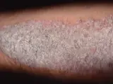
- Dermoscopy: non-specific, but useful for ruling out: fine dotted vessels on a dull-pink background with whitish streaks in between. What is decisive is what is missing — no pigment network (against a melanocytic lesion), no marginal scaling (against tinea corporis), no evenly distributed red dots with silvery scale (against psoriasis).
- Symptoms: mostly asymptomatic, occasionally with mild pruritus.
Histology
- Characteristic: palisading granuloma with histiocytes + lymphocytes (= palisading granuloma).
- Mucin in the center of the granuloma (Alcian blue +) — important for DD to rheumatoid nodules (no mucin) and Necrobiosis lipoidica (cholesterol, less mucin).
- Immunohistochemistry: CD68+ histiocytes, CD4+/CD8+ lymphocytes, few eosinophils.
Exam essentials
- Key point: annular skin-coloured to reddish plaques with a raised border WITHOUT scaling – mainly on the backs of the hands and feet.
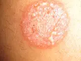
- Most important distinction: tinea corporis scales at the edge and is positive on fungal testing; granuloma annulare does not scale.
- Histology: palisading histiocytes around degenerated collagen with increased mucin (palisading granuloma).
- Forms: localised (most common), generalised/disseminated, subcutaneous, perforating.
Differential diagnoses
 Tinea corporis
 Psoriasis vulgaris (plaque psoriasis)
 Nummular eczema (discoid eczema)
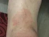 Mycosis fungoides
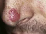 Granuloma faciale
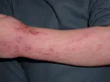 Atopic dermatitis (atopic eczema)
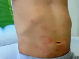 Urticaria
 Cutaneous sarcoidosis
Image credits for the thumbnails
- Tinea corporis: Corina G., “Tinea corporis”, Public Domain, via Wikimedia Commons · Public domain · Source
- Psoriasis vulgaris (plaque psoriasis): MediaJet, “An Arm Covered With Plaque Type Psoriasis”, CC BY-SA 3.0, via Wikimedia Commons · CC BY-SA 3.0 · Source
- Nummular eczema (discoid eczema): Ifnord, “Nummular dermatitits”, CC BY-SA 4.0, via Wikimedia Commons · CC BY-SA 4.0 · Source
- Mycosis fungoides: Wikindrea, “Mycosis Fungoides Before Sun and Cartamo”, CC BY-SA 4.0, via Wikimedia Commons · CC BY-SA 4.0 · Source
- Granuloma faciale: unknown (Commons user), “Facial eosinophilic granuloma 01”, CC BY 2.0, via Wikimedia Commons · CC BY 2.0 · Source
- Atopic dermatitis (atopic eczema): Eisfelder, “Atopic dermatitis child”, CC BY-SA 3.0, via Wikimedia Commons · CC BY-SA 3.0 · Source
- Urticaria: Gzzz, “Urticaria child”, CC BY-SA 4.0, via Wikimedia Commons · CC BY-SA 4.0 · Source
- Cutaneous sarcoidosis: Nowack et al., “Cutaneous findings in systemic sarcoidosis”, CC BY-SA 2.0, via Wikimedia Commons · CC BY-SA 2.0 · Source
Related conditions: how to tell them apart
- Tinea corporis – annular but with scaling at the edge – fungal testing decides
- Necrobiosis lipoidica – also a palisading granuloma, typically on the shins
- Cutaneous sarcoidosis – annular variants possible; histologically naked epithelioid granulomas
Treatment principles
- As localised granuloma annulare often regresses spontaneously, watchful waiting is also acceptable.
- Treatment is stepwise: localised disease with topical or intralesional glucocorticoids, or alternatively topical calcineurin inhibitors or cryotherapy, and generalised or refractory disease with phototherapy and systemic anti-inflammatory treatments.
- In the generalised form, screening for metabolic disorders such as diabetes mellitus and dyslipidaemia is part of the work-up.
Specific treatment regimens with drugs and doses are covered in the DermaFuchs app.
Keep learning in the app
Cross-references
Mentioned in
Keep learning
Keep learning with flashcards, exam questions and spot diagnoses in the DermaFuchs app – free, in your browser or as an app.
Related pages: Tinea corporis · Psoriasis vulgaris (plaque psoriasis) · Nummular eczema (discoid eczema)
Note: This page is intended for medical education and does not replace diagnosis or treatment decisions in individual cases.