Hypereosinophilic syndrome (HES)
- Synonyms
- HES, hypereosinophilic syndrome
- Specialty
- Dermatology · Inflammatory skin diseases
- Images
- Clinical 3
- In the app
- 1 flashcard
- Last updated
- 10/2026 · Dr. Pascal Bafteh
Contents
Images (3)
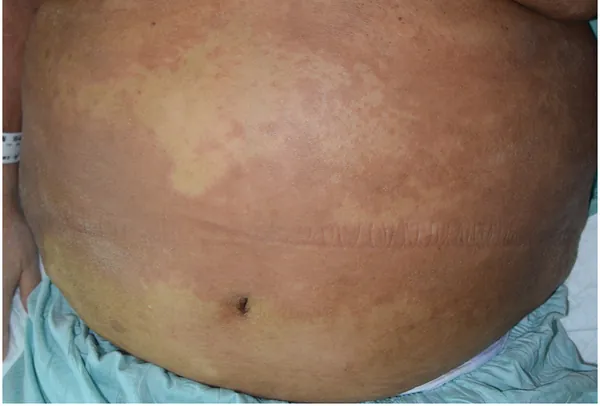
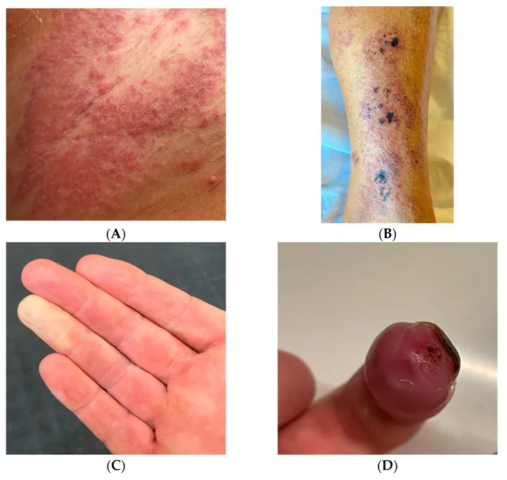
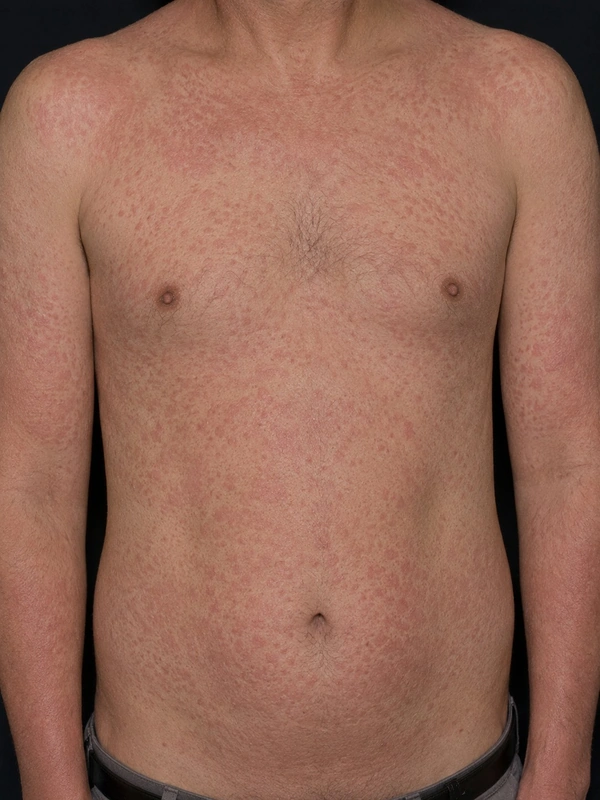 AI illustration
Definition
- Definition: HES is defined as Eosinophilia ≥ 1 500/µl on repeated counts with eosinophil-related organ involvement (heart, lung, skin, GI tract, CNS); classically persisting ≥ 6 months, today ≥ 2 counts at least 2 weeks apart suffice.
- Skin manifestations: Pruritus + urticarial plaques + erythematous papules + rarely mucosal ulcers.
Classification
- Primary (neoplastic): clonal eosinophilia in a haematological neoplasm.
- Secondary (reactive): eosinophilia in parasitic infections, allergies, drug reactions, lymphomas or other tumours.
- Idiopathic: no cause detectable.
Diagnosis
- Repeated blood counts with absolute eosinophil count, organ assessment (heart: echocardiography and troponin; lungs; nervous system).
- Exclusion of secondary causes (parasite serology, stool examination, drug history), bone marrow examination.
Differential diagnoses
 Atopic dermatitis (atopic eczema)
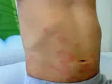 Urticaria
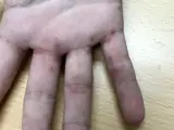 Scabies
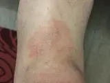 Mycosis fungoides
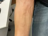 Eosinophilic fasciitis (Shulman)
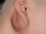 Kimura disease
Image credits for the thumbnails
- Atopic dermatitis (atopic eczema): Eisfelder, “Atopic dermatitis child”, CC BY-SA 3.0, via Wikimedia Commons · CC BY-SA 3.0 · Source
- Urticaria: Gzzz, “Urticaria child”, CC BY-SA 4.0, via Wikimedia Commons · CC BY-SA 4.0 · Source
- Scabies: Gzzz, “Scabies hand and fingers 1”, CC BY-SA 4.0, via Wikimedia Commons · CC BY-SA 4.0 · Source
- Mycosis fungoides: Wikindrea, “Mycosis Fungoides Before Sun and Cartamo”, CC BY-SA 4.0, via Wikimedia Commons · CC BY-SA 4.0 · Source
- Eosinophilic fasciitis (Shulman): Matthews C, Thomas D, Monteagudo L et al. (Clin Case Rep 2025), CC BY 4.0, via PubMed Central (PMC11926566) · CC BY 4.0 · Source
- Kimura disease: Zhang G et al. (BMC Surg 2020), CC BY 4.0, via PubMed Central (PMC6941305) · CC BY 4.0 · Source
Treatment principles
- Management is multidisciplinary, usually led by haematology, and depends on the subtype and organ involvement; secondary eosinophilia, for example due to parasites, drugs or allergies, is excluded first.
- In the idiopathic and lymphocytic variants, systemic glucocorticoids are first line, with biologics targeting interleukin-5 or other immunosuppressants added as steroid-sparing agents; before high-dose glucocorticoids, Strongyloides infection is excluded or treated, as it can otherwise cause life-threatening disseminated infection.
- The myeloproliferative variant with FIP1L1-PDGFRA fusion responds very well to tyrosine kinase inhibitors.
Specific treatment regimens with drugs and doses are covered in the DermaFuchs app.
Keep learning in the app
References (selection)
- Contemporary consensus proposal on criteria and classification of eosinophilic disorders and related syndromes
- A tyrosine kinase created by fusion of the PDGFRA and FIP1L1 genes as a therapeutic target of imatinib in idiopathic hypereosinophilic syndrome
- DermNet: Hypereosinophilic syndrome
- MSD Manual Professional: Hypereosinophilic syndrome
- Hypereosinophilia: clinical and therapeutic approach in 2025
Cross-references
Keep learning
Keep learning with flashcards, exam questions and spot diagnoses in the DermaFuchs app – free, in your browser or as an app.
Related pages: Atopic dermatitis (atopic eczema) · Urticaria · Scabies
Note: This page is intended for medical education and does not replace diagnosis or treatment decisions in individual cases.