Kimura disease
- Specialty
- Dermatology · Inflammatory skin diseases
- Images
- Histology 1 · Clinical 2
- In the app
- 2 flashcards
- Last updated
- 10/2026 · Dr. Pascal Bafteh
Contents
Images (3)
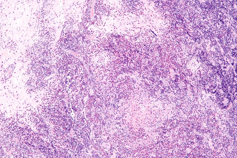 Histology
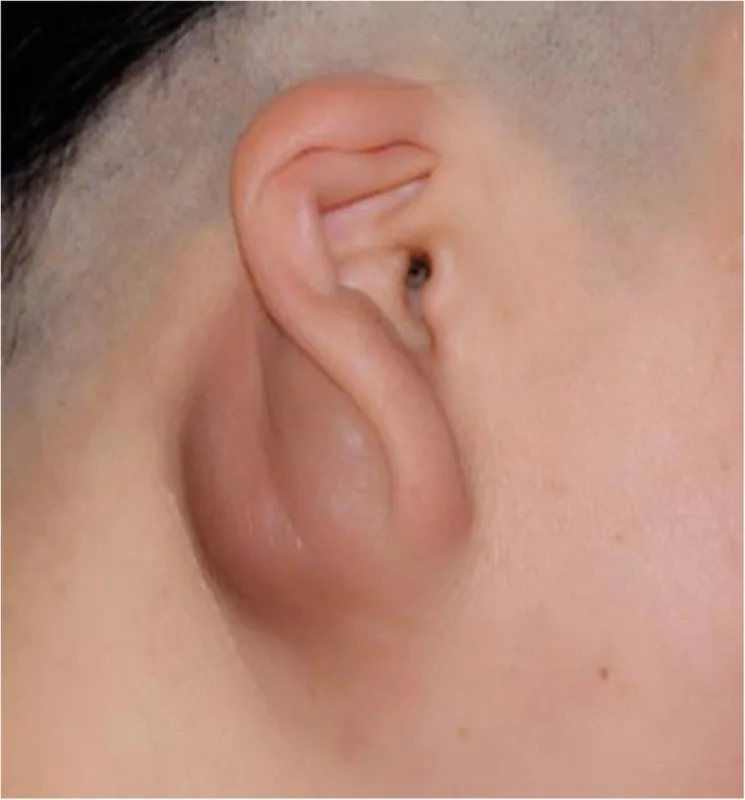
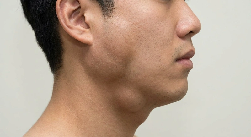 AI illustration
Definition
Kimura disease is a rare chronic inflammatory disease of unknown cause with painless subcutaneous swellings and enlarged lymph nodes in the head and neck region, blood eosinophilia and raised serum IgE; it mainly affects young men of East Asian heritage.
Diagnosis
- Biopsy: reactive follicular hyperplasia, eosinophil-rich infiltrate, fibrosis and arborising vascular proliferation.
- Blood count (eosinophilia), total IgE, urinalysis and kidney function (proteinuria, nephrotic syndrome).
- The key differential is angiolymphoid hyperplasia with eosinophilia (more superficial, less often with lymphadenopathy, eosinophilia and raised IgE); also lymphomas.
Differential diagnoses
 Angioedema
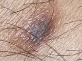 Dermatofibroma
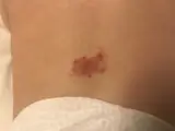 Haemangioma
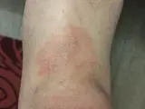 Mycosis fungoides
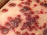 Kaposi's sarcoma
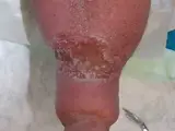 Lymphoedema
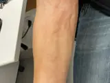 Eosinophilic fasciitis (Shulman)
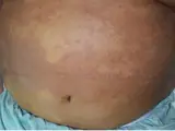 Hypereosinophilic syndrome (HES)
Image credits for the thumbnails
- Angioedema: James Heilman, MD, “AngioedemaFra”, CC BY-SA 3.0, via Wikimedia Commons · CC BY-SA 3.0 · Source
- Dermatofibroma: Jmarchn, “Pigmented and tenderness dermatofibroma”, CC BY-SA 3.0, via Wikimedia Commons · CC BY-SA 3.0 · Source
- Haemangioma: Gstk, “Infantile hemangioma, flat”, CC BY-SA 4.0, via Wikimedia Commons · CC BY-SA 4.0 · Source
- Mycosis fungoides: Wikindrea, “Mycosis Fungoides Before Sun and Cartamo”, CC BY-SA 4.0, via Wikimedia Commons · CC BY-SA 4.0 · Source
- Kaposi's sarcoma: unknown author, “Kaposi's Sarcoma”, Public Domain, via Wikimedia Commons · Public Domain · Source
- Lymphoedema: Wesalius, “Lymphedema̠ right leg.jpg”, CC BY-SA 4.0, via Wikimedia Commons · CC BY-SA 4.0 · Source
- Eosinophilic fasciitis (Shulman): Matthews C, Thomas D, Monteagudo L et al. (Clin Case Rep 2025), CC BY 4.0, via PubMed Central (PMC11926566) · CC BY 4.0 · Source
- Hypereosinophilic syndrome (HES): Hida Y, Hara T (JAAD Case Rep 2026), CC BY 4.0, via PubMed Central (PMC13280059) – cropped (abdomen) · CC BY 4.0 · Source
Treatment principles
- Surgical excision is the treatment of choice for circumscribed lesions; recurrence is common.
- Alternatively, or in recurrence, intralesional or systemic glucocorticoids, other immunosuppressants and radiotherapy may be considered.
- As renal involvement, particularly nephrotic syndrome, can occur, monitoring of urine and renal function is part of care.
More on this topic in the DermaFuchs app.
Keep learning in the app
References (selection)
Cross-references
Keep learning
Keep learning with flashcards, exam questions and spot diagnoses in the DermaFuchs app – free, in your browser or as an app.
Related pages: Angioedema · Dermatofibroma · Haemangioma
Note: This page is intended for medical education and does not replace diagnosis or treatment decisions in individual cases.