Cutaneous sarcoidosis
Exam relevance: in 3 of more than 50 board exam reports · rank 45
- Synonyms
- sarcoidosis
- Specialty
- Dermatology · Inflammatory skin diseases
- Images
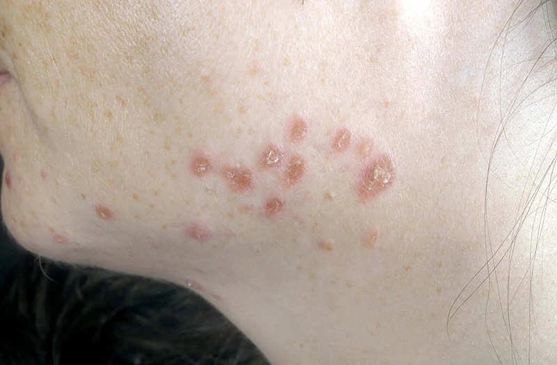
- Clinical 1 · Histology 1
- In the app
- 10 flashcards
- Last updated
- 10/2026 · Dr. Pascal Bafteh
Contents
Images (2)
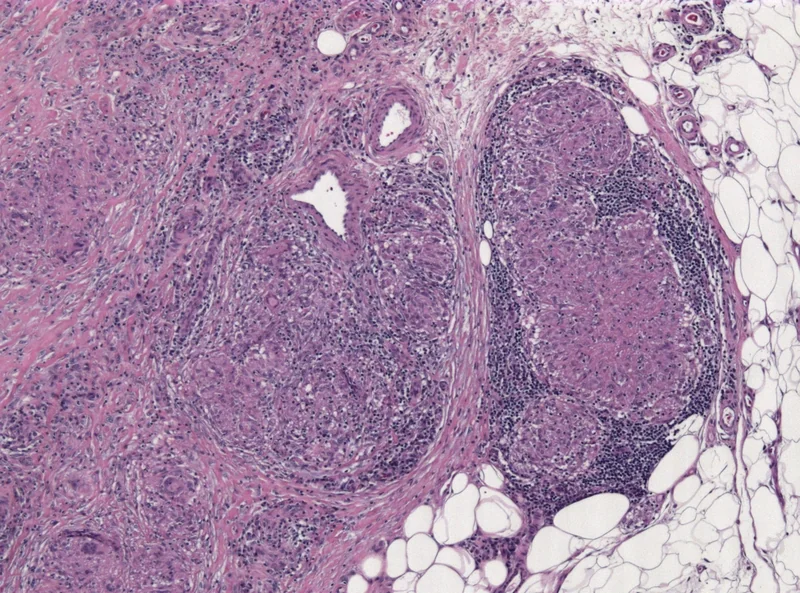 Histology
Definition
- Epidemiology: F: M ≈ 2: 1, more common in African Americans and Scandinavians. First manifestation 20–40 years.
- Pathogenesis Hypothesis: Misdirection by Antigen presentation of infectious triggers with CD4 T-cells + IL-2 + ACE.
- Clinical Features: Multiorgan involvement possible — Skin + lungs + eyes + kidneys (lungs > 90 %, often bilateral hilar lymphadenopathy).
- Skin findings: lupoid plaques (“Lupus pernio”), macules, annular / psoriasiform / ichthyosiform variants, scar sarcoidosis in tattoos / scars.
- Lupus pernio: livid-red to bluish-violet, doughy infiltrated plaques on the nose, cheeks, earlobes and fingers — the hallmark of chronic persistent sarcoidosis, frequently associated with involvement of the nasal mucosa and upper airways and with fibrosing pulmonary disease; prognostically unfavourable and often refractory (MTX, hydroxychloroquine, anti-TNF in reserve — exclude TB beforehand). Despite the name it has no relationship to the chilblain lupus of lupus erythematosus.
| Feature | Lupus pernio | Rosacea |
|---|---|---|
| Colour | livid-bluish, "chilblain-like" | bright red to coppery red |
| Telangiectasias | absent | characteristically present |
| Papulopustules | none | typical |
| Surface | doughy infiltrated, smooth plaque | papules/pustules, bulbous in the phyma stage |
| Diascopy (glass spatula) | apple-jelly colour of the granuloma | erythema blanches |
| Histology | non-caseating epithelioid granulomas | perifollicular inflammation; granulomas only in granulomatous rosacea |
| Systemic signs | lungs, eyes, raised sIL-2R and ACE | none |
| Flush/triggers | none | heat, alcohol, spicy food |
- Special Forms:
- Heerfordt's Syndrome (Uveoparotid fever): Uveitis + parotitis + facial nerve palsy ± fever — cranial nerve involvement typical.
- Diagnostics: Chest X-ray + sIL-2R + ACE + BAL (CD4/CD8 ↑); biopsy with non-caseating granulomas.
Differential diagnoses
 Lupus erythematosus
 Rosacea
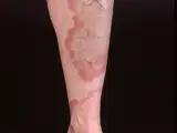 Granuloma annulare
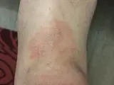 Mycosis fungoides
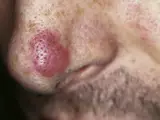 Granuloma faciale
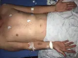 Secondary syphilis (lues II)
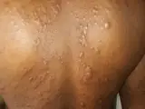 Lepromatous leprosy
- Lupus Pernio (Sarcoidosis)
Image credits for the thumbnails
- Lupus erythematosus: Meriam Hajji, CC BY 4.0, via Wikimedia Commons · CC BY 4.0 · Source
- Rosacea: Michael Sand, Daniel Sand, Christina Thrandorf, Volker Paech, Peter Altmeyer, Falk G Bechara, “Rosacea”, CC BY 2.5, via Wikimedia Commons, cropped · CC BY 2.5 · Source
- Granuloma annulare: Kreuter et al., “Generalized granuloma annulare”, CC BY-SA 2.0, via Wikimedia Commons · CC BY-SA 2.0 · Source
- Mycosis fungoides: Wikindrea, “Mycosis Fungoides Before Sun and Cartamo”, CC BY-SA 4.0, via Wikimedia Commons · CC BY-SA 4.0 · Source
- Granuloma faciale: unknown (Commons user), “Facial eosinophilic granuloma 01”, CC BY 2.0, via Wikimedia Commons · CC BY 2.0 · Source
- Secondary syphilis (lues II): Ehlers S, Sergent S, Ashurst J (Clin Pract Cases Emerg Med 2020), CC BY 4.0, via PubMed Central (PMC7676796) · CC BY 4.0 · Source
- Lepromatous leprosy: Shiyanbola AC, Emezie PC, Saheed O et al. (Cureus 2026), CC BY 4.0, via PubMed Central (PMC13294642) · CC BY 4.0 · Source
Treatment principles
- Systemic sarcoidosis must be investigated before treatment; if organs are involved, management is interdisciplinary.
- Circumscribed skin lesions are treated with potent topical or intralesional glucocorticoids.
- Extensive, disfiguring or refractory forms, such as lupus pernio, warrant systemic therapy, e.g. with antimalarials, systemic glucocorticoids, other immunosuppressants or TNF inhibitors.
Specific treatment regimens with drugs and doses are covered in the DermaFuchs app.
Keep learning in the app
References (selection)
Cross-references
Mentioned in
Keep learning
Keep learning with flashcards, exam questions and spot diagnoses in the DermaFuchs app – free, in your browser or as an app.
Related pages: Lupus erythematosus · Rosacea · Granuloma annulare
Note: This page is intended for medical education and does not replace diagnosis or treatment decisions in individual cases.