Graft-versus-host disease (GvHD)
- Synonyms
- GvHD, graft-versus-host disease
- Specialty
- Dermatology · Inflammatory skin diseases
- Images
- Clinical 2
- In the app
- 2 flashcards
- Last updated
- 10/2026 · Dr. Pascal Bafteh
Contents
Images (2)
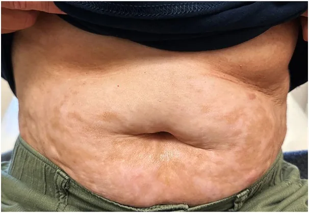
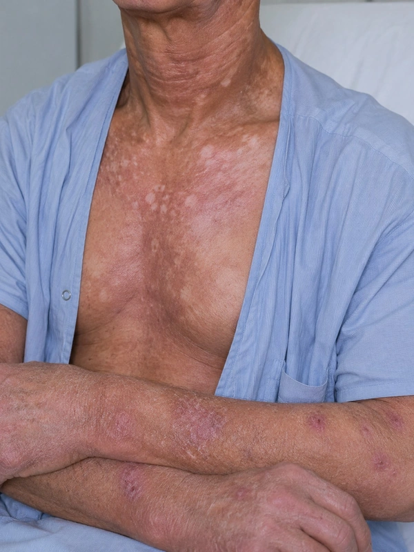 AI illustration
Definition
Definition & Epidemiology
- GvHD = Donor immune cells recognize recipient antigens as foreign → systemic immune reaction after allogeneic stem cell or organ transplantation due to differences in HLA and minor histocompatibility antigens (hence also with an HLA-identical donor).
- The skin is the most common and earliest organ involved in acute GvHD.
Aetiopathogenesis
Pathogenesis — 3 Phases (Ferrara-Model)
- Phase 2 — Activation: Donor T-cells recognise recipient alloantigens and expand clonally.
- Phase 3 — Effector phase: Donor T-cells attack via FasL + perforin/granzyme + TNF-α/IFN-γ.
- Chronic GvHD (separate model, not one of the 3 phases): Th17 / Th2 shift + fibroblast activation → Scleroderma-like skin changes and fibrosis of internal organs.
Acute GvHD (aGvHD, < 100 days post-Tx)
- Clinical presentation: Initially maculopapular rash on acral areas/ear helix/cheeks/neck (DD: AME!), later confluent to erythrodermic.
- Severity stages (Glucksberg / Mount Sinai):
- Stage 1: Rash < 25% BSA
- Stage 2: 25–50%
- Stage 3: > 50%, generalized
- Stage 4: Erythroderma + bullae / epidermolysis (TEN-like!) — prognostically unfavorable
- Extra-cutaneous: Hepatitis (transaminases / bilirubin ↑), GvHD enteritis (secretory diarrhea, abdominal pain, bloody stools), conjunctivitis.
Chronic GvHD (cGvHD, > 100 days post-Tx)
- NIH criteria 2014 with diagnostic and distinctive manifestations.
- Skin manifestations — 3 course types:
- Lichenoid cGvHD: Shows lichen planus-like papules + Wickham striae plus reticular oral mucosa — DD: Lichen planus.
- Scleroderma-like cGvHD: diffuse skin sclerosis, possibly fascial involvement, nail changes, pterygium unguium.
- Poikilodermatous presentation: Hyper-/hypopigmentation + atrophy + telangiectasias — DD: Mycosis fungoides poikilodermatica.
- Mucositis: xerostomic / lichenoid oral GvHD, Sicca syndrome (eye, mouth, vagina), reticular mucosal changes.
- Other organs: Lungs (BOS, bronchiolitis obliterans), liver (cholestatic), GI, eyes (Sicca).
Diagnosis
- Clinical presentation + timing (acute < 100 d, chronic > 100 d).
- Histology (punch biopsy, perilesional): Apoptotic keratinocytes in basal layer, vacuolization, lymphocytic infiltrate ("satellite cell necrosis"), in scleroderma-like cGvHD thickened dermis + fibrosis.
- Differential diagnoses: Drug reaction (DRESS, maculopapular AME), viral exanthem, toxic shock, engraftment syndrome.
Differential diagnoses
 Limited systemic sclerosis (CREST syndrome)
 Lupus erythematosus
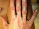 Dermatomyositis
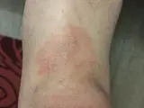 Mycosis fungoides
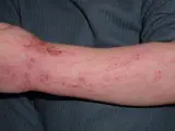 Atopic dermatitis (atopic eczema)
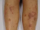 Lichen planus
 Psoriasis vulgaris (plaque psoriasis)
 Lichen sclerosus
Image credits for the thumbnails
- Limited systemic sclerosis (CREST syndrome): James Heilman, MD, “CREST syndrome (calcinosis and sclerodactyly)”, CC BY-SA 4.0, via Wikimedia Commons · CC BY-SA 4.0 · Source
- Lupus erythematosus: Meriam Hajji, CC BY 4.0, via Wikimedia Commons · CC BY 4.0 · Source
- Dermatomyositis: Mohammad2018, CC BY-SA 4.0 · CC BY-SA 4.0 · Source
- Mycosis fungoides: Wikindrea, “Mycosis Fungoides Before Sun and Cartamo”, CC BY-SA 4.0, via Wikimedia Commons · CC BY-SA 4.0 · Source
- Atopic dermatitis (atopic eczema): Eisfelder, “Atopic dermatitis child”, CC BY-SA 3.0, via Wikimedia Commons · CC BY-SA 3.0 · Source
- Lichen planus: James Heilman, MD, “Lichen Planus (2)”, CC BY-SA 3.0, via Wikimedia Commons · CC BY-SA 3.0 · Source
- Psoriasis vulgaris (plaque psoriasis): MediaJet, “An Arm Covered With Plaque Type Psoriasis”, CC BY-SA 3.0, via Wikimedia Commons · CC BY-SA 3.0 · Source
- Lichen sclerosus: Frucht CS, Ragsdale BD, Cureus 2026, Figure 1, CC BY 4.0, via PubMed Central (PMC13614091) · CC BY 4.0 · Source
Treatment principles
- Treatment is coordinated closely with the transplant centre.
- First-line treatment is systemic glucocorticoids with optimised calcineurin inhibitor prophylaxis; in steroid-refractory disease, JAK inhibitors, extracorporeal photopheresis or other immunosuppressants are used.
- Cutaneous manifestations are additionally treated with topical glucocorticoids or calcineurin inhibitors and phototherapy, and sclerodermatous forms with accompanying physiotherapy to prevent contractures.
Specific treatment regimens with drugs and doses are covered in the DermaFuchs app.
Keep learning in the app
Cross-references
Keep learning: test yourself
One question from the DermaFuchs app on Graft-versus-host disease (GvHD) – tap an answer.
Question 1 of 1
Which clinical feature completes the triad of poikiloderma alongside hyper-/hypopigmentation and telangiectasias in Graft-versus-Host-Disease?
Answer
Correct: atrophy. Poikiloderma is defined by the triad of hyper- and hypopigmentation, telangiectasias, and epidermal atrophy.
More questions like these – with explanations and spaced repetition – in the app.
Note: This page is intended for medical education and does not replace diagnosis or treatment decisions in individual cases.