Psoriasis vulgaris (plaque psoriasis)
Definition
Chronic inflammatory Th17-mediated dermatosis with histological hyperproliferation and parakeratosis of the epidermis. Characteristic is a "antifungal-defense state" of the skin with overreaction of the IL-23/IL-17 axis to external triggers.
Brief Etiopathogenesis
Immunology: Th17 inflammation with IL-17A/F, IL-23 and TNF-α. Genetics: predisposition via PSORS1–15 loci, especially HLA-Cw6. Triggers: mechanical stimuli and infections (esp. streptococci). Pathology: accelerated epidermal regeneration (transit time ↓ from 30 days to 5–8 days).
Main Forms
Plaque psoriasis (psoriasis vulgaris, 80–90%), guttate psoriasis, pustular psoriasis, palmoplantar psoriasis, inverse psoriasis, erythrodermic psoriasis, nail psoriasis, psoriatic arthritis.
Clinical Findings and Phenomena
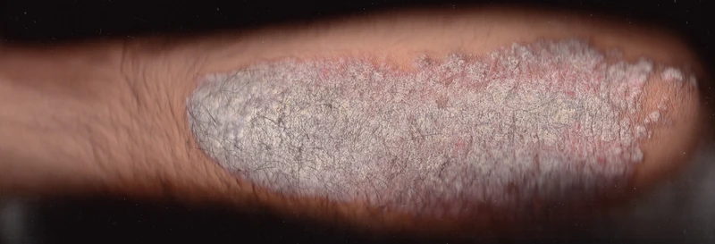
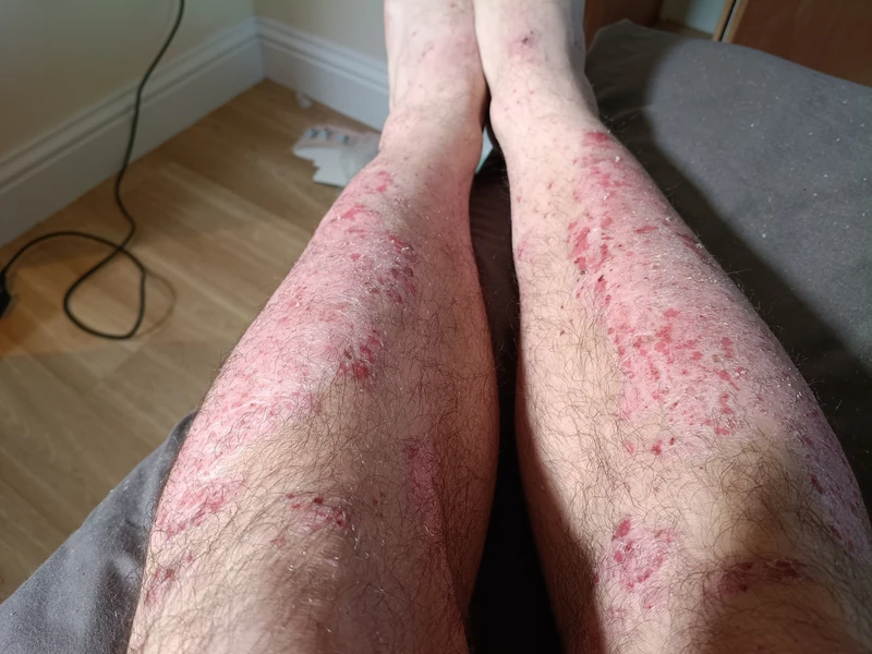
Typical skin lesions are sharply demarcated, silvery-white scaling erythemato-squamous plaques.
- Koebner phenomenon: new psoriasis lesions on previously healthy skin after mechanical trauma (scratching, pressure, injury).
- Candle-wax phenomenon: scale can be scraped off like wax.
- Last membrane sign: further scraping reveals a thin intact epithelial layer.
- Auspitz phenomenon: removing this layer produces pinpoint bleeding from injured capillary loops (papillomatosis).
Epidemiology — Comparative Figures for the Common Inflammatory Dermatoses
In Europe psoriasis affects about 2–3 % of the population — clearly more common than lichen planus and less common than atopic dermatitis in childhood.
| Disease | Prevalence | Comment |
|---|---|---|
| Psoriasis vulgaris | approx. 2–3 % of adults | two peaks of manifestation (see below) |
| Atopic dermatitis | children approx. 10–20 %, adults approx. 2–5 % | most common chronic skin disease of childhood |
| Lichen planus | approx. 0.5–1 % of adults | peak in middle adulthood |
| Psoriatic arthritis | 6–40 % of psoriasis patients | see section 5 |
- Two peaks of manifestation in psoriasis: type I (early onset) before the age of 40, strongly HLA-Cw6-associated, positive family history, usually more severe; type II (late onset) after the age of 40, less often familial, milder course.
- Men and women are affected with about equal frequency.
Dermoscopy
Dotted/glomerular vessels on erythematous background — uniform dot pattern vs. seborrheic dermatitis with branching vessels.
Histopathology
Hyper- and parakeratosis, absent stratum granulosum, acanthosis and papillomatosis. Elongated, thin rete ridges, club-shaped papillary bodies. Sub- or intracorneal Munro microabscesses. Diffuse lymphocytic infiltrate in the dermis with histiocytes, CD4+ lymphocytes and neutrophils.
Pustular form: dense accumulations of intact neutrophils spongiformly traversing the epidermis (Kogoj pustule).
Basic Diagnostic Work-up (clinical + triggers)
- Diagnosis is primarily clinical, based on typical morphology and distribution. Biopsy only in unclear cases.
- Streptococcal serology (especially in guttate psoriasis): anti-streptolysin O, anti-DNase B, anti-hyaluronidase.
- Swabs from tonsils, anal rim, nasopharynx.
- Rheumatoid factor and anti-CCP to exclude rheumatoid arthritis.
- Mycological diagnostics to exclude dermatophytosis.
Definition and Basic Principle
Psoriasis vulgaris is a chronic inflammatory, T-cell-mediated autoimmune skin disease which may also affect joints, entheses and internal organs. It is based on immune dysregulation in a genetically predisposed individual, activated by external trigger factors (e.g. infections, stress, medications or mechanical stimuli).
Genetics and Epidemiology
Type I: onset usually before age 40 (peak 2nd–3rd decade), strongly associated with HLA-C06:02, familial clustering, often more severe. Type II: onset after age 40 (5th–6th decade), usually no HLA-C06:02, tends to be milder.
In Europe, 2–3 % prevalence; bimodal age distribution. Nail involvement in 40–50 %, psoriatic arthritis in ~33 %.
Systemic Associations & Comorbidities
Psoriasis is now considered a multisystem inflammatory disease with relevance beyond the skin. Associated: obesity & metabolic syndrome, cardiovascular disease, Crohn's and uveitis, depression and psychiatric disease, nicotine/alcohol dependence, liver fibrosis (MASLD).
Mutual exclusion: atopic eczema and allergic contact eczema less common in psoriasis.
Differential diagnoses
- Seborrhoeic dermatitis
- Tinea corporis
- Pityriasis rosea
- Guttate psoriasis
- Atopic dermatitis (atopic eczema)
- Nummular eczema
- Pityriasis versicolor
- Pityriasis rubra pilaris
Practise Psoriasis vulgaris (plaque psoriasis) in the app
Flashcards with spaced repetition, exam questions and spot-the-diagnosis on this topic – in DermaFuchs, free of charge.
In the DermaFuchs app: 11 flashcards · 20 clinical images
Open in browser Download on the App StoreSources (selection)
- Marrakchi et al. 2022
- AWMF S3 Psoriasis 2021
- Marghoob 2019 Dermoscopy Review
- De Rosa et al. 2007
- Chen et al. 2018
- Jackson et al. 2024
More inflammatory skin diseases
- Cheilitis granulomatosa (Miescher)
- Erythema annulare centrifugum (EAC)
- Erythema nodosum
- Graft-versus-host disease (GvHD)
- Granuloma annulare
- Cutaneous sarcoidosis
- Lichen planus
- Lichen sclerosus
Note: This page is intended for medical education and does not replace diagnosis or treatment decisions in individual cases. Treatment and follow-up content is available in the app.