Erythema nodosum
Exam relevance: in 2 of more than 50 board exam reports · rank 69
- Synonyms
- tender red nodules on shins, Löfgren syndrome
- Specialty
- Dermatology · Inflammatory skin diseases
- Images
- Clinical 4
- In the app
- 7 flashcards
- Last updated
- 10/2026 · Dr. Pascal Bafteh
Contents
Images (4)
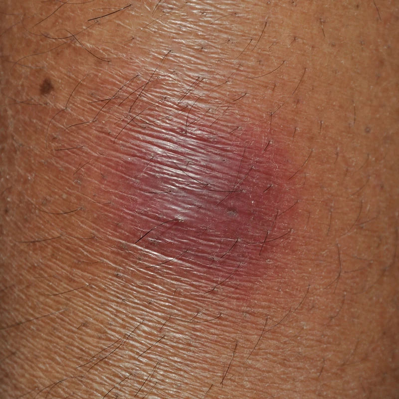
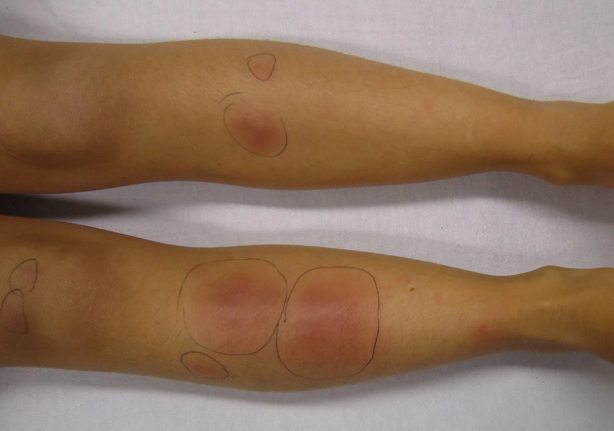
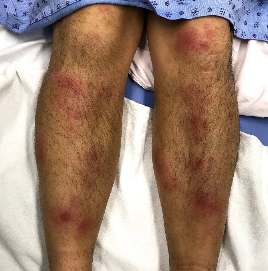
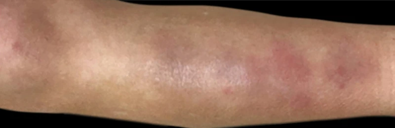
Definition
Erythema nodosum is the most common form of panniculitis: a reactive inflammatory disorder of the subcutaneous fat (septal panniculitis without vasculitis) triggered by various infections, drugs and systemic diseases, or remaining idiopathic.
- Epidemiology: Women 20–40 years, self-limiting over 3–6 weeks.
- Clinical Features: painful reddish-livid subcutaneous nodules on anterior shins, no ulcer, healing with hyperpigmentation without scarring. Fever + arthralgias + general symptoms.
- Trigger Spectrum: Classically Streptococci + TB + Sarcoidosis plus Crohn's disease + pregnancy + oral contraceptives.
- Histology: septal panniculitis without vasculitis; Miescher's radial granulomas = characteristic but not pathognomonic star-shaped histiocytic granulomas around a central cleft (also in erythema induratum, Sweet syndrome, Behçet disease).
Clinical features
- Acutely appearing, very tender, poorly defined, bright red to violaceous nodules, symmetrically on the shins, less often on knees, thighs or forearms. Sources disagree on size: a review gives 1–5 cm (Leung et al. 2018, children), DermNet NZ 3–20 cm.
- Colour changes like a bruise over time (red-purple, green-yellow), no ulceration, no scarring.
- Often accompanied by fever, fatigue and arthralgia; in Löfgren syndrome (acute sarcoidosis) the chest X-ray shows bilateral hilar lymphadenopathy.
Diagnosis
- Usually a clinical diagnosis; search for the trigger: history (infections, drugs including hormonal contraception, pregnancy, bowel symptoms).
- Targeted search for the common triggers: streptococcal infection (throat swab, antistreptolysin titre), tuberculosis and sarcoidosis (chest X-ray), inflammatory bowel disease, drugs and pregnancy.
- Biopsy (deep spindle-shaped biopsy into the fat) only in atypical cases.
Differential diagnoses
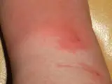 Erysipelas
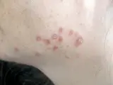 Cutaneous sarcoidosis
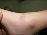 Polyarteritis nodosa
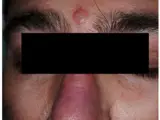 Sweet syndrome (acute febrile neutrophilic dermatosis)
- Löfgren's Syndrome / Acute Sarcoidosis
Image credits for the thumbnails
- Erysipelas: Poupou l'quourouce, “Erysipel”, CC BY-SA 3.0, via Wikimedia Commons · CC BY-SA 3.0 · Source
- Cutaneous sarcoidosis: Nowack et al., “Cutaneous findings in systemic sarcoidosis”, CC BY-SA 2.0, via Wikimedia Commons · CC BY-SA 2.0 · Source
- Polyarteritis nodosa: Ruby Haviv, Maya Capua, Jacob Amir, Liora Harel, “Cutaneous polyarteritis nodosa”, CC BY 4.0, via Wikimedia Commons · CC BY 4.0 · Source
- Sweet syndrome (acute febrile neutrophilic dermatosis): Antonelli E et al. (J Clin Med 2021), CC BY 4.0, via PubMed Central (PMC7835974) · CC BY 4.0 · Source
Treatment principles
- Erythema nodosum usually resolves spontaneously within weeks; the key is to identify and treat the underlying cause, such as streptococcal infection, sarcoidosis, inflammatory bowel disease or a causative drug.
- Rest, leg elevation and compression as well as non-steroidal anti-inflammatory drugs provide symptomatic relief.
- In severe cases, systemic glucocorticoids may be used once infection has been excluded.
Specific treatment regimens with drugs and doses are covered in the DermaFuchs app.
Keep learning in the app
References (selection)
Cross-references
Mentioned in
Keep learning: test yourself
Two questions from the DermaFuchs app on Erythema nodosum – tap an answer.
Question 1 of 2
What type of panniculitis is associated with erythema nodosum?
Answer
Correct: Septal panniculitis. Erythema nodosum is characterized by septal panniculitis, which is inflammation of the septa between fat lobules.
Question 2 of 2
Where do the painful reddish-livid subcutaneous nodules of erythema nodosum typically appear?
Answer
Correct: anterior shins. Erythema nodosum classically presents as painful, red subcutaneous nodules on the anterior shins.
More questions like these – with explanations and spaced repetition – in the app.
Note: This page is intended for medical education and does not replace diagnosis or treatment decisions in individual cases.