Sporotrichosis
- Synonyms
- rose gardener's disease
- Specialty
- Dermatology · Bacterial infections
- Images
- Clinical 2
- In the app
- 4 flashcards
- Last updated
- 10/2026 · Dr. Pascal Bafteh
Contents
Images (2)
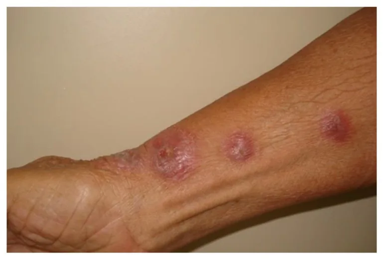
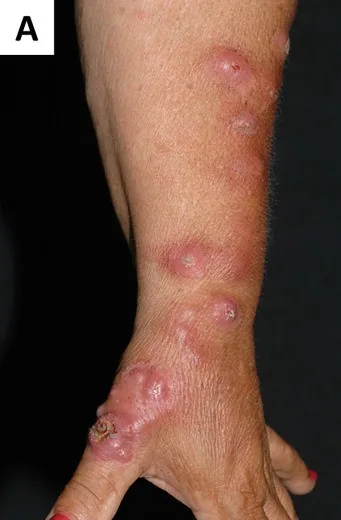
Definition
- Pathogen is Sporothrix schenckii, a dimorphic fungus with hyphae at 25 °C, yeast at 37 °C — in gardeners after minor injuries from plant thorns.
- Manifestation weeks-months after injury on hands/forearms with bluish-red nodules/plaques and a tendency to ulceration.
- Sporotrichoid arrangement: linear ascending nodules along lymphatic vessels — important differential diagnosis to M. marinum (same pattern!).
- Clinical features: papulonodular lesions with lymphangitic spread ("sporotrichoid course")
- Transmission: roses, soil
- Differential diagnoses: atypical mycobacteria, nocardiosis
Differential diagnoses
 Mycosis fungoides
 Cutaneous leishmaniasis
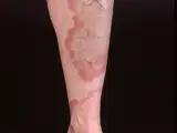 Granuloma annulare
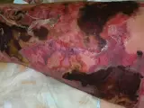 Necrotizing fasciitis
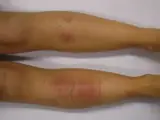 Erythema nodosum
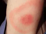 Erythema migrans (Lyme disease)
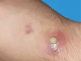 Swimming Pool Granuloma
Image credits for the thumbnails
- Mycosis fungoides: Wikindrea, “Mycosis Fungoides Before Sun and Cartamo”, CC BY-SA 4.0, via Wikimedia Commons · CC BY-SA 4.0 · Source
- Cutaneous leishmaniasis: Abanima, “Cutaneous leishmaniasis2”, CC BY-SA 3.0, via Wikimedia Commons · CC BY-SA 3.0 · Source
- Granuloma annulare: Kreuter et al., “Generalized granuloma annulare”, CC BY-SA 2.0, via Wikimedia Commons · CC BY-SA 2.0 · Source
- Necrotizing fasciitis: CNX OpenStax, CC BY 4.0, via Wikimedia Commons, cropped · CC BY 4.0 · Source
- Erythema nodosum: James Heilman, MD, “ENlegs”, CC BY-SA 3.0, via Wikimedia Commons · CC BY-SA 3.0 · Source
- Erythema migrans (Lyme disease): James Gathany (CDC), “Erythema migrans - erythematous rash in Lyme disease - PHIL 9875”, Public Domain, via Wikimedia Commons · Public domain · Source · CDC PHIL #9875
- Swimming Pool Granuloma: Kyriazidis I, Trakatelli M, Spyropoulou GA., "When the Usual Becomes Unusual: A Closer Look at Mycobacterium marinum Infections", Cureus 2024, Figure 3, CC BY 4.0, via PMC · CC BY 4.0 · Source
Treatment principles
- Sporotrichosis is treated with systemic antifungals, usually for several months and beyond clinical healing.
- Disseminated or extracutaneous disease, especially in immunosuppression, requires more intensive, initially intravenous antifungal therapy.
- Small, localised lesions can be treated additionally by surgical or physical methods.
Specific treatment regimens with drugs and doses are covered in the DermaFuchs app.
Keep learning in the app
Cross-references
Keep learning: test yourself
One question from the DermaFuchs app on Sporotrichosis – tap an answer.
Question 1 of 1
What is the primary clinical morphology of cutaneous sporotrichosis lesions before they ulcerate?
Answer
Correct: bluish-red nodules/plaques. Sporotrichosis initially presents as bluish-red nodules or plaques that often progress to ulceration.
More questions like these – with explanations and spaced repetition – in the app.
Note: This page is intended for medical education and does not replace diagnosis or treatment decisions in individual cases.