Relapsing polychondritis
- Specialty
- Dermatology · Connective tissue diseases
- Images
- Clinical 2
- In the app
- 2 flashcards
- Last updated
- 10/2026 · Dr. Pascal Bafteh
Contents
Images (2)
Definition
Definition & Epidemiology
- Relapsing polychondritis (relapsing polychondritis, RP) — rare autoimmune episodic cartilage inflammation that can affect all cartilaginous structures of the body (ear cartilage, nasal septum, trachea, larynx, joints, eyes, heart valves).
- Epidemiology: Incidence ~3–5/million/year; peak 40–60 years of age; no sex predilection.
- Associations: ~30% combined with other autoimmune diseases (RA, SLE, Sjögren, vasculitides, Myelodysplastic Syndrome — VEXAS syndrome!).
Aetiopathogenesis
Pathogenesis
- Autoimmune reaction against Type II collagen + Matrilin-1.
- Genetic: associated with HLA-DR4 and – as a separate entity – the acquired somatic UBA1 mutation of VEXAS syndrome (mainly middle-aged and older men).
Auricular Chondritis
(most common finding, up to 90%)
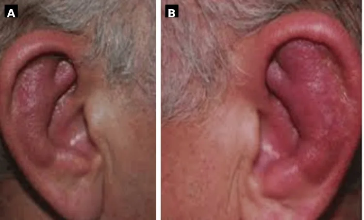
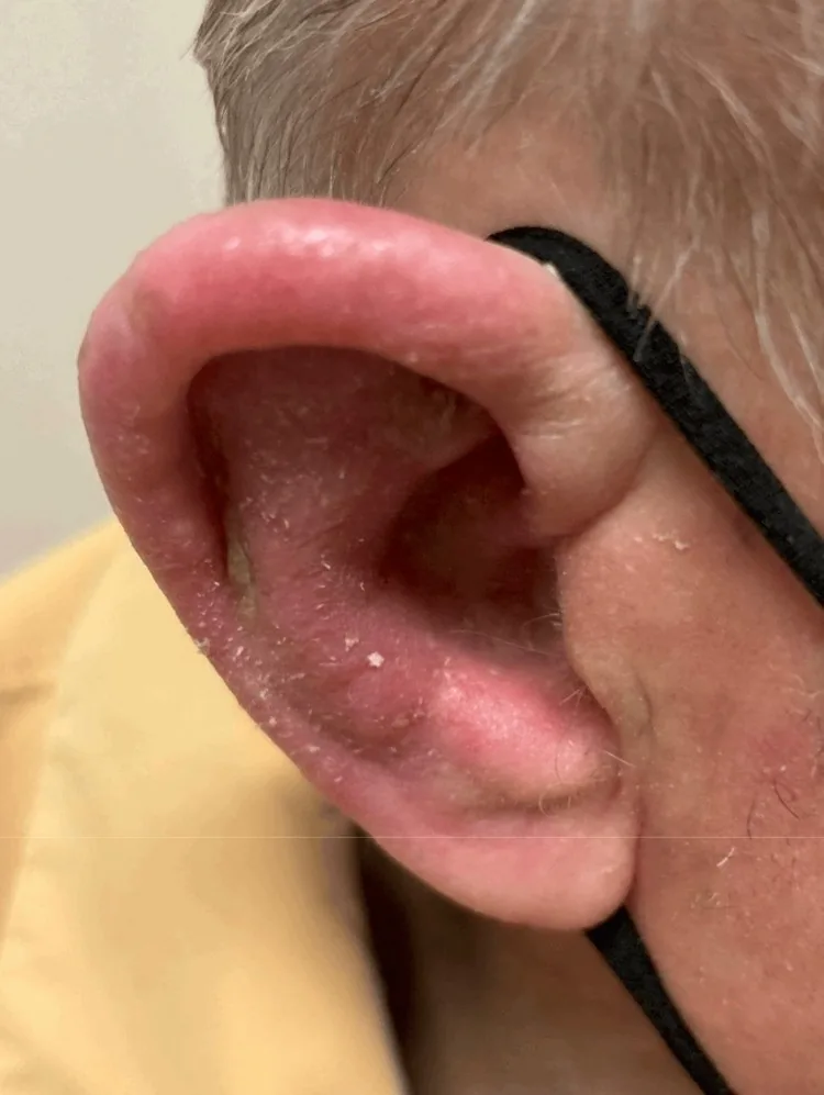
- Acute, painful livid-red, swollen auricle with sparing of the lobule — lobule sparing (no cartilage) is classic.
- Repeated flares → "Cauliflower Ear" due to cartilage destruction.
Tracheobronchial Involvement
(~50%, life-threatening!)
- Hoarseness, cough, stridor, dyspnea; collapsing airway → emergency.
- A major cause of death in RP is respiratory complication.
Diagnosis
Diagnostic Criteria (McAdam, modified by Damiani)
- 1: Bilateral auricular chondritis
- 2: Non-erosive seronegative inflammatory polyarthritis
- 3: Nasal chondritis
- 4: Ocular involvement
- 5: Airway cartilage involvement
- 6: Audiovestibular damage
Diagnostics
- Laboratory: ↑ ESR, CRP; Anti-Type II collagen antibodies (50%, not specific); ANA, ANCA for DD.
- Imaging: HRCT thorax (tracheal wall thickening, stenosis), echocardiography.
- Histology of ear biopsy: perichondrial lymphocyte-plasma cell infiltrate.
- Exclude DD VEXAS: UBA1 mutation in men > 50 years with MDS/refractory disease.
Differential diagnoses
 Erysipelas
 Angioedema
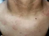 Lupus erythematosus
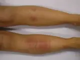 Erythema nodosum
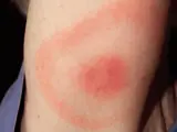 Erythema migrans (Lyme disease)
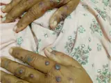 Granulomatosis with polyangiitis (GPA) – skin manifestations
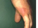 Erysipeloid
Image credits for the thumbnails
- Erysipelas: Poupou l'quourouce, “Erysipel”, CC BY-SA 3.0, via Wikimedia Commons · CC BY-SA 3.0 · Source
- Angioedema: James Heilman, MD, “AngioedemaFra”, CC BY-SA 3.0, via Wikimedia Commons · CC BY-SA 3.0 · Source
- Lupus erythematosus: Meriam Hajji, CC BY 4.0, via Wikimedia Commons · CC BY 4.0 · Source
- Erythema nodosum: James Heilman, MD, “ENlegs”, CC BY-SA 3.0, via Wikimedia Commons · CC BY-SA 3.0 · Source
- Erythema migrans (Lyme disease): James Gathany (CDC), “Erythema migrans - erythematous rash in Lyme disease - PHIL 9875”, Public Domain, via Wikimedia Commons · Public domain · Source · CDC PHIL #9875
- Granulomatosis with polyangiitis (GPA) – skin manifestations: Ullah N, Jabin A, Isar H et al. (Ann Med Surg (Lond) 2026), CC BY 4.0, via PubMed Central (PMC13461037) · CC BY 4.0 · Source
- Erysipeloid: GP MARTINEAU, “Rouget humain”, CC BY-SA 3.0, via Wikimedia Commons · CC BY-SA 3.0 · Source · cropped
Treatment principles
- Treatment depends on organ involvement: mild flares are treated with non-steroidal anti-inflammatory drugs and systemic glucocorticoids.
- Involvement of the airways, eyes or heart requires high-dose glucocorticoids and steroid-sparing immunosuppressants, with biologics for refractory disease.
- Care is multidisciplinary; airway stenosis may require stenting or a tracheostomy.
Specific treatment regimens with drugs and doses are covered in the DermaFuchs app.
Keep learning in the app
Cross-references
Keep learning
Keep learning with flashcards, exam questions and spot diagnoses in the DermaFuchs app – free, in your browser or as an app.
Related pages: Erysipelas · Angioedema · Lupus erythematosus
Note: This page is intended for medical education and does not replace diagnosis or treatment decisions in individual cases.