Leukocytoclastic vasculitis
Overview
- Mechanism: Classic small-vessel vasculitis (leukocytoclastic vasculitis) is a type III reaction according to Coombs and Gell — an immune complex reaction.
- Sequence: soluble antigen-antibody complexes are deposited, with slight antigen excess, in the wall of postcapillary venules — predilection for the lower legs (hemodynamics/stasis).
- Complement activation: activation of the complement system via the classical pathway with release of the anaphylatoxins C3a and C5a; C5a is chemotactic for neutrophil granulocytes.
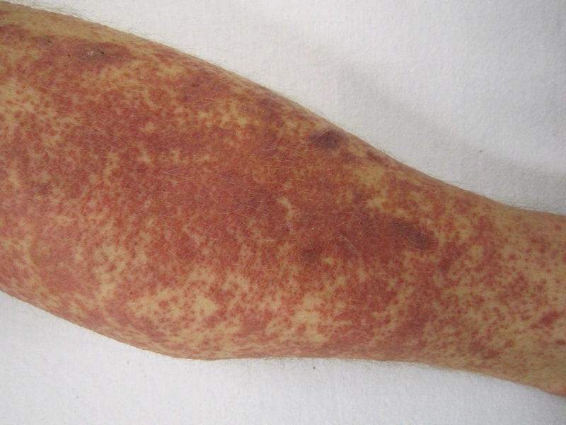
- Tissue damage: the neutrophils release lysosomal enzymes and reactive oxygen species → fibrinoid necrosis of the vessel wall and leukocytoclasia (nuclear dust) as the histological correlate; extravasation of erythrocytes explains the palpable, non-blanching purpura.
- Laboratory correlate: complement consumption with low C3 and/or C4 — particularly marked in hypocomplementemic urticarial vasculitis and cryoglobulinemic vasculitis; low complement argues for an immune-complex-mediated process.
- Direct immunofluorescence: biopsy from a fresh lesion (< 24–48 h) — perivascular Ig and C3 deposits; IgA deposits characterize IgA vasculitis (Henoch-Schönlein purpura).
- Counter-check: The ANCA-associated vasculitides (GPA, EGPA, MPA) are not immune-complex-mediated but pauci-immune — DIF remains largely negative and complement is normal.
- Model reaction of type III = Arthus reaction.
Overview
- Histology: Neutrophilic infiltrate + nuclear dust. Clinical presentation: Palpable purpura on lower extremities.
- Procedure: Biopsy (< 48 hours after onset), DIF, Labs (CBC, CRP, Crea, Urine, Complement, ANCA, ANA, Cryoglobulins, Hepatitis serology).
- Pathomechanism: Type III immune complex reaction — deposition of circulating immune complexes in postcapillary venules, activation of the complement system (C3a/C5a) and consecutive recruitment of neutrophils.
- Triggers: Infections (classically a preceding streptococcal throat infection), drugs (e.g. beta-lactam antibiotics, NSAIDs), chronic viral hepatitis B/C (cryoglobulinaemic vasculitis), autoimmune disease and malignancy; a substantial proportion of cases remains idiopathic.
- DIF specimen: Direct immunofluorescence requires a second, unfixed biopsy (transport in saline or Michel’s medium, processed as a frozen section) — formalin destroys the immunoreactants.
Differential diagnoses
- Erythema nodosum
- Polyarteritis nodosa
- Purpura fulminans
- Erythema migrans (Lyme disease)
- IgA vasculitis (Henoch-Schönlein purpura)
- Urticaria
- Urticarial vasculitis
Practise Leukocytoclastic vasculitis in the app
Flashcards with spaced repetition, exam questions and spot-the-diagnosis on this topic – in DermaFuchs, free of charge.
In the DermaFuchs app: 8 flashcards · 5 clinical images
Open in browser Download on the App StoreSources (selection)
- Plewig G, French L, Ruzicka T, Kaufmann R, Hertl M (Hrsg). Braun-Falcos Dermatologie, Venerologie und Allergologie. 7. Aufl. Berlin: Springer; 2018.
- Delaleu et al. 2022
More vascular skin disorders
- Erythromelalgia
- Lichen aureus
- Superficial thrombophlebitis
- Great saphenous vein varicosity
- Deep vein thrombosis
Note: This page is intended for medical education and does not replace diagnosis or treatment decisions in individual cases. Treatment and follow-up content is available in the app.