Majocchi granuloma
- Synonyms
- deep dermatophytosis
- Specialty
- Dermatology · Fungal infections
- Images
- Clinical 1
- In the app
- 1 flashcard
- Last updated
- 10/2026 · Dr. Pascal Bafteh
Contents
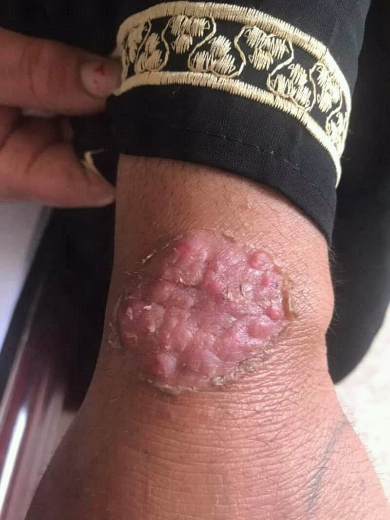
Images (1)
Definition
Majocchi granuloma is a deep follicular dermatophyte infection in which fungi reach the dermis through ruptured hair follicles and cause granulomatous inflammation there. The most common pathogen is Trichophyton rubrum.
- Clinical features: firm follicular nodules and pustular papules with deep dermal component — inflammatory dermal dermatophytosis, often on legs after shaving.
- Pathogenesis: Seeding of dermatophytes (mostly T. rubrum) along follicularly ruptured structures deep into the dermis.
- Diagnostics: Punch biopsy with PAS staining (hyphae perifollicularly in dermis).
Occurrence & epidemiology
It mainly affects adults. Predisposing factors are a longstanding, persistent dermatophyte infection, shaving or other skin trauma, a tropical climate and immunosuppression (e.g. after organ transplantation); the deep nodular form occurs mainly in immunosuppression.
Differential diagnoses
 Tinea corporis
 Furuncle and carbuncle
 Erythema nodosum
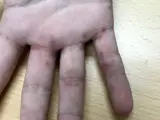 Scabies
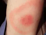 Erythema migrans (Lyme disease)
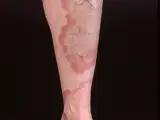 Granuloma annulare
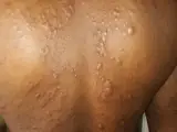 Lepromatous leprosy
Image credits for the thumbnails
- Tinea corporis: Corina G., “Tinea corporis”, Public Domain, via Wikimedia Commons · Public domain · Source
- Furuncle and carbuncle: Medicalpal, “Carbuncle”, CC BY-SA 4.0, via Wikimedia Commons · CC BY-SA 4.0 · Source
- Erythema nodosum: James Heilman, MD, “ENlegs”, CC BY-SA 3.0, via Wikimedia Commons · CC BY-SA 3.0 · Source
- Scabies: Gzzz, “Scabies hand and fingers 1”, CC BY-SA 4.0, via Wikimedia Commons · CC BY-SA 4.0 · Source
- Erythema migrans (Lyme disease): James Gathany (CDC), “Erythema migrans - erythematous rash in Lyme disease - PHIL 9875”, Public Domain, via Wikimedia Commons · Public domain · Source · CDC PHIL #9875
- Granuloma annulare: Kreuter et al., “Generalized granuloma annulare”, CC BY-SA 2.0, via Wikimedia Commons · CC BY-SA 2.0 · Source
- Lepromatous leprosy: Shiyanbola AC, Emezie PC, Saheed O et al. (Cureus 2026), CC BY 4.0, via PubMed Central (PMC13294642) · CC BY 4.0 · Source
Treatment principles
- Because the dermatophytes have penetrated deep into the follicles and dermis, topical treatment alone is insufficient; systemic antifungal therapy over several weeks is required.
- The pathogen should first be confirmed by microscopy, culture or molecular methods.
- Topical glucocorticoids, which often act as a trigger, should be stopped; topical antimycotics can be used as an adjunct.
Guideline: AWMF guideline register (no. 013-002)
Specific treatment regimens with drugs and doses are covered in the DermaFuchs app.
Keep learning in the app
References (selection)
Cross-references
Keep learning
Keep learning with flashcards, exam questions and spot diagnoses in the DermaFuchs app – free, in your browser or as an app.
Related pages: Tinea corporis · Furuncle and carbuncle · Erythema nodosum
Note: This page is intended for medical education and does not replace diagnosis or treatment decisions in individual cases.