Contact dermatitis (contact eczema)
Exam relevance: in 9 of more than 50 board exam reports · rank 12
- Synonyms
- Kontaktdermatitis, Kontaktallergie, allergisches Kontaktekzem, Nickelallergie, irritatives Kontaktekzem, contact dermatitis, contact allergy, nickel allergy, allergic contact dermatitis, irritant contact dermatitis
- Specialty
- Dermatology · Allergies
- Images
- Clinical 4 · Histology 1
- In the app
- 9 flashcards
- Last updated
- 10/2026 · Dr. Pascal Bafteh
Contents
Images (5)
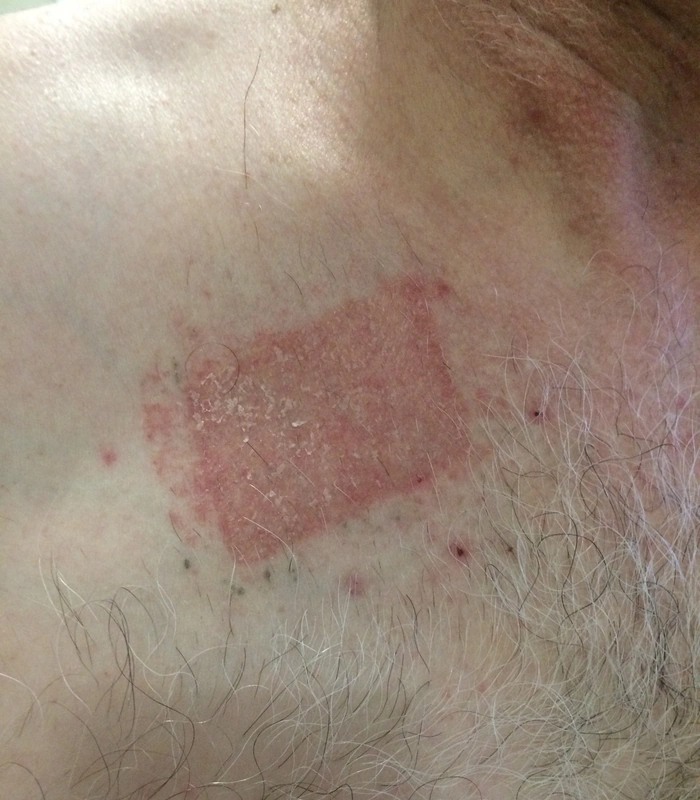
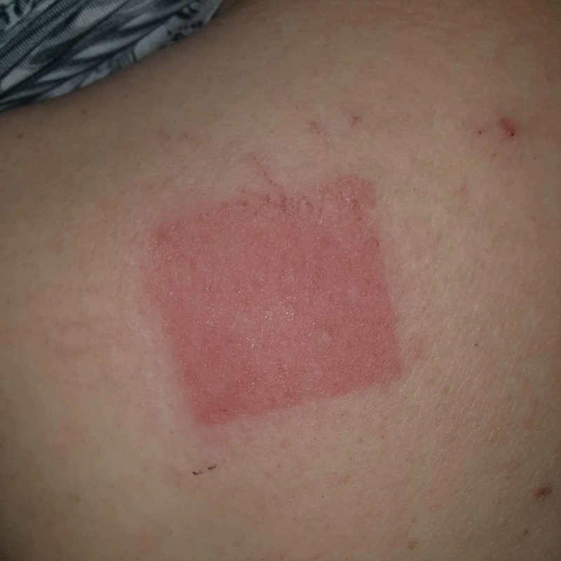
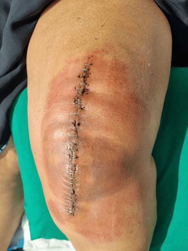
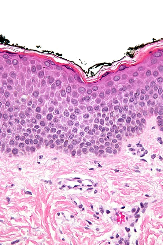 Histology
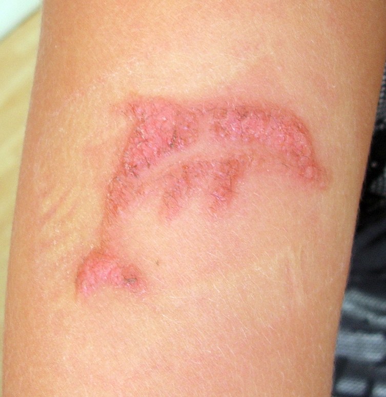
Definition
Contact dermatitis (contact eczema) is an inflammatory skin reaction at the site where an external substance touches the skin. Allergic contact dermatitis is a T-cell-mediated delayed-type reaction (Coombs and Gell type IV) in previously sensitised individuals; irritant contact dermatitis develops without sensitisation through direct damage to the skin barrier.
Classification
- Allergic contact dermatitis: sensitisation may occur even after years of symptom-free contact; thereafter eczema typically appears 24–72 hours after renewed contact, even with minute amounts; spreading beyond the contact area is possible.
- Irritant contact dermatitis: acute after a single strong noxa (acids, alkalis) or chronic-cumulative after repeated weak insults such as wet work, detergents and friction; usually confined to the exposed area.
- Special forms: photoallergic contact dermatitis (allergen plus UV light), airborne contact dermatitis (e.g. plant particles) and protein contact dermatitis.
Occurrence & epidemiology
Contact dermatitis is the most common cause of occupational skin disease; about 80% of occupational hand dermatitis is irritant. Wet-work occupations such as cleaning, healthcare, food handling/catering and hairdressing, as well as construction, are particularly affected. Common contact allergens in Europe include nickel (the most common allergen), fragrances, preservatives (e.g. methylchloro-/methylisothiazolinone), balsam of Peru, colophony, p-phenylenediamine (hair dyes), chromate, cobalt and rubber chemicals (thiurams).
Aetiopathogenesis
In allergic contact dermatitis, low-molecular-weight haptens penetrate the skin, bind to body proteins and are carried by Langerhans cells and dermal dendritic cells to the regional lymph nodes. Hapten-specific T cells develop there and trigger the eczematous reaction on renewed contact. In irritant dermatitis, detergents, solvents or prolonged moisture directly damage the stratum corneum lipids and keratinocytes. A pre-existing barrier defect, for example in atopic skin, favours both forms.
Clinical features
- Acute: erythema, oedema, papulovesicles to blisters, oozing and crusts with intense itch.
- Chronic: scaling, lichenification, hyperkeratosis and painful fissures, typically on the hands.
- Distribution: morphology and site are often the best clues to the cause – e.g. nickel at contact sites of jewellery, piercings or a watch strap, p-phenylenediamine in hair dye with swelling and blistering of the face and neck, methylisothiazolinone from hair products or wet wipes on the face, colophony under adhesive plasters; allergens transferred by the fingers may also cause dermatitis of the eyelids or genitals.
- Allergic contact dermatitis may spread beyond the contact area, whereas irritant dermatitis tends to stay sharply confined to the exposed zone.
Histology
Spongiotic dermatitis with intercellular epidermal oedema, spongiotic vesicles and a perivascular lymphocytic infiltrate, sometimes with eosinophils; in chronic disease acanthosis and parakeratosis. In allergic contact dermatitis eosinophils may be prominent in the epidermis (eosinophilic spongiosis); irritant contact dermatitis tends to show mild spongiosis, keratinocyte necrosis and neutrophils in the epidermis.
Diagnosis
- History: time course, occupation, hobbies, cosmetics, jewellery and the distribution pattern.
- Patch test: the gold standard for allergic contact dermatitis. Diluted test substances (standard series plus occupational or product-related series) are taped onto the back under occlusion for 48 hours; readings are taken when the patches come off and again a few days later, because reactions usually take 48–96 hours to develop. Testing is done on healed, calm skin.
- Irritant contact dermatitis is a diagnosis of exclusion with matching exposure and a negative patch test.
Differential diagnoses
 Atopic dermatitis (atopic eczema)
 Seborrhoeic dermatitis
 Dyshidrotic eczema (pompholyx)
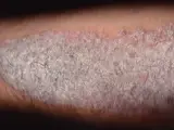 Psoriasis vulgaris (plaque psoriasis)
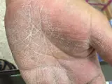 Tinea manuum
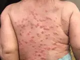 Eczema herpeticum
 Erythrasma
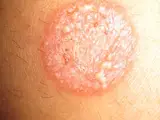 Tinea corporis
Image credits for the thumbnails
- Atopic dermatitis (atopic eczema): Eisfelder, “Atopic dermatitis child”, CC BY-SA 3.0, via Wikimedia Commons · CC BY-SA 3.0 · Source
- Seborrhoeic dermatitis: Roymishali, “Seborrhoeic dermatitis highres”, CC BY-SA 3.0, via Wikimedia Commons · CC BY-SA 3.0 · Source
- Dyshidrotic eczema (pompholyx): Inwe (Wikimedia Commons), “Dyshidrosis”, CC BY-SA 3.0, via Wikimedia Commons · CC BY-SA 3.0 · Source
- Psoriasis vulgaris (plaque psoriasis): MediaJet, “An Arm Covered With Plaque Type Psoriasis”, CC BY-SA 3.0, via Wikimedia Commons · CC BY-SA 3.0 · Source
- Tinea manuum: Mohammad2018, “Tinea manum”, CC BY-SA 4.0, via Wikimedia Commons · CC BY-SA 4.0 · Source
- Eczema herpeticum: Mohammad2018, CC BY-SA 4.0, via Wikimedia Commons, cropped · CC BY-SA 4.0 · Source
- Erythrasma: unknown (Commons user), “Erythrasma”, CC BY-SA 4.0, via Wikimedia Commons · CC BY-SA 4.0 · Source
- Tinea corporis: Corina G., “Tinea corporis”, Public Domain, via Wikimedia Commons · Public domain · Source
Treatment principles
- The key step is identifying and consistently avoiding the trigger; in allergic contact dermatitis this includes patch testing.
- Acute lesions are treated with topical glucocorticoids, accompanied by emollient care and skin protection; extensive disease may require short-term systemic glucocorticoids.
- In chronic hand eczema, phototherapy and systemic retinoids may be considered; if the cause is occupational, the occupational dermatology reporting procedure (Hautarztverfahren) should be initiated.
Specific treatment regimens with drugs and doses are covered in the DermaFuchs app.
Keep learning in the app
References (selection)
- DermNet: Contact dermatitis
- DermNet: Allergic contact dermatitis
- DermNet: Irritant contact dermatitis
- DermNet: Patch tests
- DermNet: Eczema pathology
- The European baseline series in 10 European Countries, 2005/2006--results of the European Surveillance System on Contact Allergies (ESSCA)
- Immunopathogenesis and immunoregulatory mechanisms in allergic contact dermatitis
Cross-references
Mentioned in
Keep learning: test yourself
One question from the DermaFuchs app on Contact dermatitis (contact eczema) – tap an answer.
Question 1 of 1
What is the underlying pathomechanism of allergic contact dermatitis?
Answer
Correct: Type IV delayed reaction. Allergic contact dermatitis is a Type IV delayed hypersensitivity reaction mediated by T-cells, which typically manifests 48-72 hours after allergen exposure.
More questions like these – with explanations and spaced repetition – in the app.
Note: This page is intended for medical education and does not replace diagnosis or treatment decisions in individual cases.