Extramammary Paget disease
Exam relevance: in 3 of more than 50 board exam reports · rank 45
- Synonyms
- EMPD, extramammary Paget's disease
- Specialty
- Dermatology · Skin tumours
- Images
- Clinical 2 · Histology 4
- In the app
- 4 flashcards
- Last updated
- 10/2026 · Dr. Pascal Bafteh
Contents
Images (6)
 Histology
 Histology
 Histology
 Histology
Definition
Extramammary Paget's Disease (EMPD)
Definition & Pathology
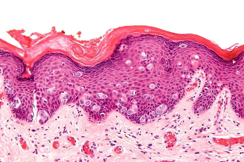
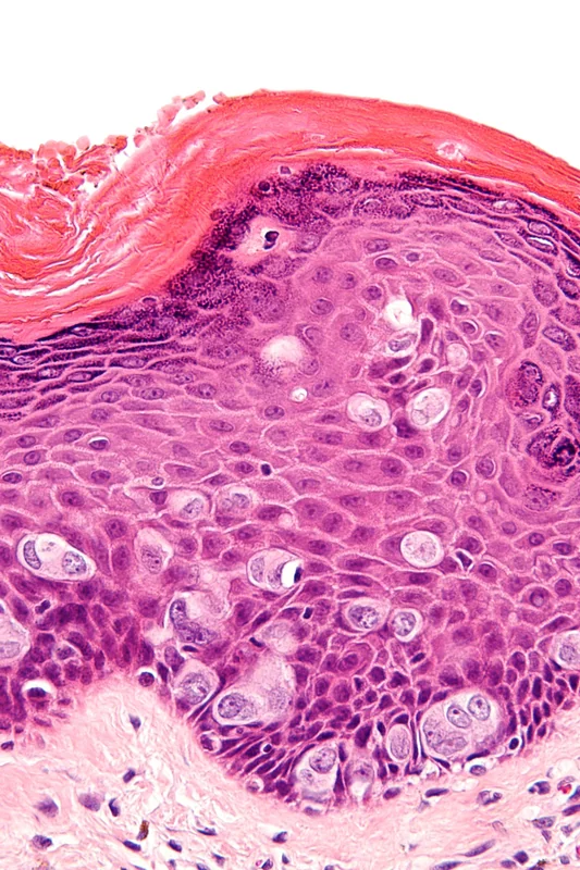
- EMPD = apocrine-differentiated intraepithelial adenocarcinoma with slow pagetoid spread in the epidermis.
- Patients: Women > Men (in Asia, however, men > women) — peak age around 65 years.
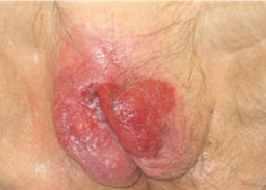
- Location: apocrine areas (anogenital + axillary) — also perineal, inguinal, periumbilical.
Clinical Presentation
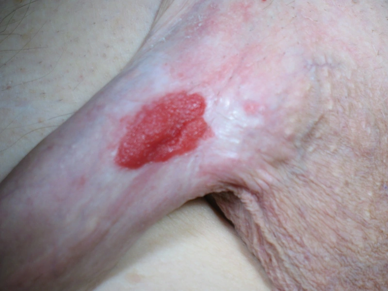
- Solitary, irregularly demarcated erosive-weeping plaque with a finely scaling surface, usually itchy.
- Common misdiagnoses: Eczema + Inverse Psoriasis — months of delay.
Histology & IHC
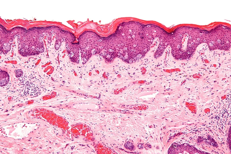
- "Pagetoid clear cells" intraepithelial (also deeper in adnexal structures).
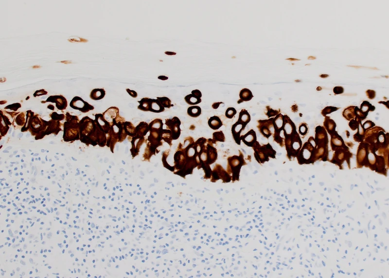
- IHC: CK7+ + CEA+ + GCDFP-15+ — Differentiation from SCC (CK7−) and melanoma (S100+, CK7−).
- Subclassification: primary cutaneous (intraepithelial) vs. secondary as spread of an internal adenocarcinoma (rectum, urothelium, breast) into the skin.
Diagnostics / Staging
- Punch biopsy + mapping biopsies for extent determination (often much larger than clinically visible!).
- Exclude secondary adenocarcinoma:
- Anogenital / Perianal: Rectoscopy + colonoscopy (in case of perianal involvement)
- Penis / Scrotum: Urogenital diagnostics (cystoscopy, ultrasound)
- Inguinal / Vulva: Gynecology + Urology consultation
- Mammography for axillary localization
- CT / Ultrasound of lymph node stations for metastasis search.
Differential diagnoses
 Bowen's disease (squamous cell carcinoma in situ)
 Psoriasis vulgaris (plaque psoriasis)
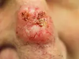 Basal cell carcinoma
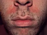 Seborrhoeic dermatitis
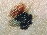 Melanoma
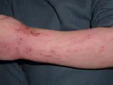 Atopic dermatitis (atopic eczema)
 Contact dermatitis (contact eczema)
Image credits for the thumbnails
- Bowen's disease (squamous cell carcinoma in situ): Crop by Mikael Häggström, from original by Klaus D. Peter, Gummersbach, Germany, “Bowen's disease (crop)”, CC BY 3.0 de, via Wikimedia Commons · CC BY 3.0 de · Source
- Psoriasis vulgaris (plaque psoriasis): MediaJet, “An Arm Covered With Plaque Type Psoriasis”, CC BY-SA 3.0, via Wikimedia Commons · CC BY-SA 3.0 · Source
- Basal cell carcinoma: Bin im Garten, “Basaliom am Nasenrücken 2”, CC BY-SA 3.0, via Wikimedia Commons · CC BY-SA 3.0 · Source
- Seborrhoeic dermatitis: Roymishali, “Seborrhoeic dermatitis highres”, CC BY-SA 3.0, via Wikimedia Commons · CC BY-SA 3.0 · Source
- Melanoma: National Cancer Institute (unknown photographer), “Melanoma (4)”, Public Domain, via Wikimedia Commons · Public domain · Source
- Atopic dermatitis (atopic eczema): Eisfelder, “Atopic dermatitis child”, CC BY-SA 3.0, via Wikimedia Commons · CC BY-SA 3.0 · Source
- Contact dermatitis (contact eczema): Dr.khatmando, “Severe contact dermatitis buprenorphine transdermal patch”, CC BY-SA 4.0, via Wikimedia Commons · CC BY-SA 4.0 · Source
Treatment principles
- The treatment of choice is micrographically controlled surgery; because the extent is often clinically underestimated, preoperative mapping biopsies are performed.
- In case of contraindications or multifocal disease, topical treatments, photodynamic therapy or radiotherapy are options, although topical treatments reach only the intraepidermal component.
- As secondary Paget disease can reflect an underlying carcinoma, this is sought and, if present, treated first.
Specific treatment regimens with drugs and doses are covered in the DermaFuchs app.
Keep learning in the app
Cross-references
Keep learning: test yourself
One question from the DermaFuchs app on Extramammary Paget disease – tap an answer.
Question 1 of 1
Which immunohistochemical profile is characteristic of Extramammary Paget's Disease?
Answer
Correct: CK7+ + CEA+ + GCDFP-15+. EMPD cells characteristically express CK7, CEA, and GCDFP-15, distinguishing them from squamous cell carcinoma and melanoma.
More questions like these – with explanations and spaced repetition – in the app.
Note: This page is intended for medical education and does not replace diagnosis or treatment decisions in individual cases.