Tinea corporis
- Synonyms
- ringworm, body ringworm
- Specialty
- Dermatology · Fungal infections
- Images
- Clinical 13
- In the app
- 11 flashcards
- Last updated
- 10/2026 · Dr. Pascal Bafteh
Contents
Images (13)
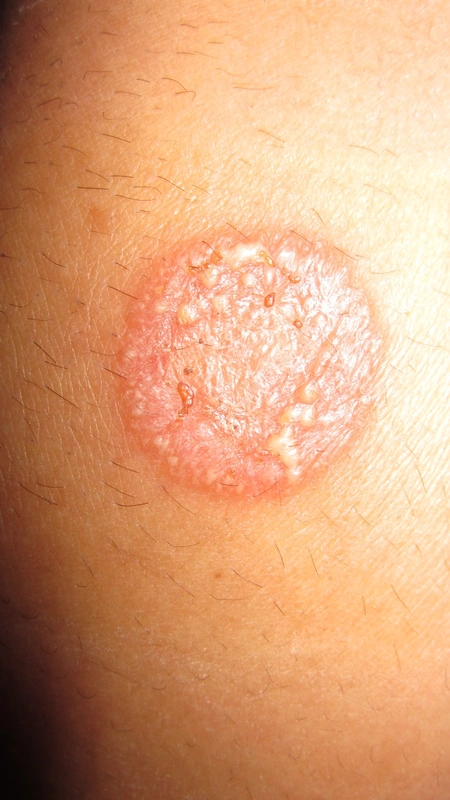
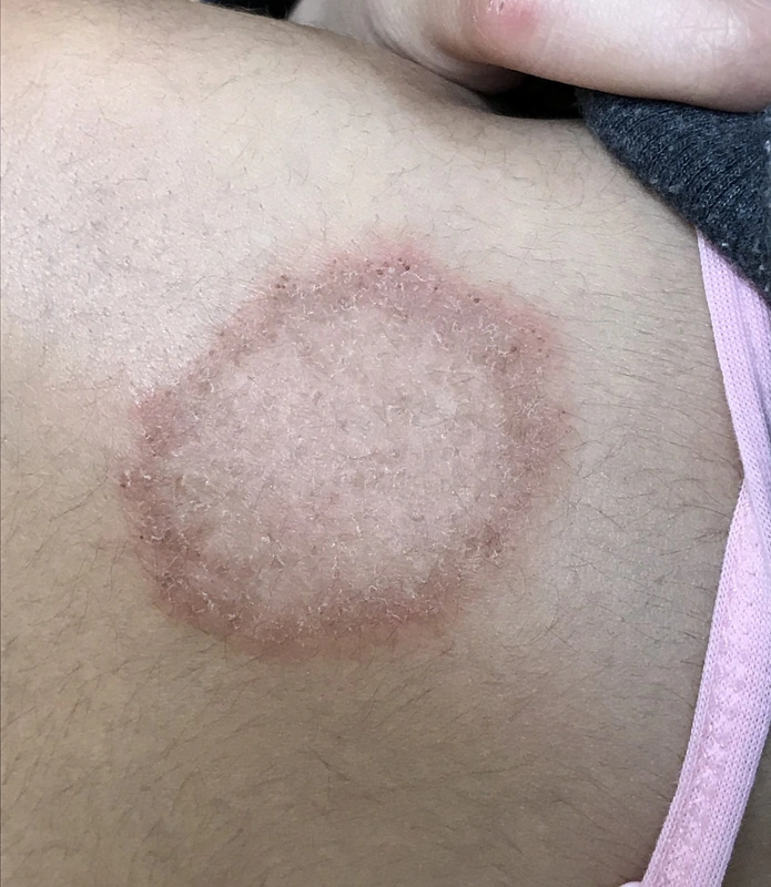
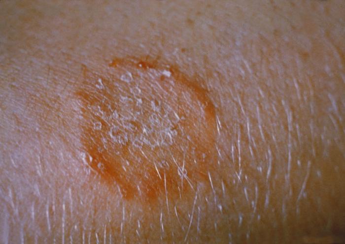
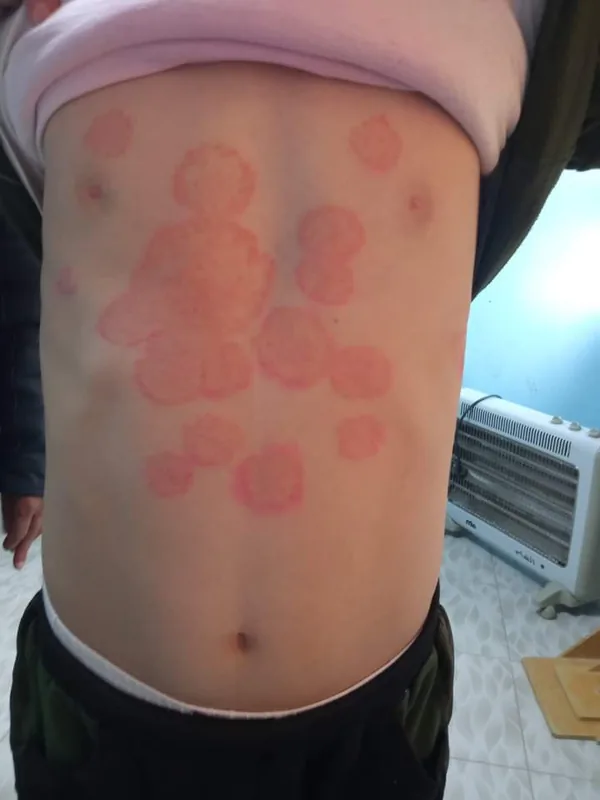
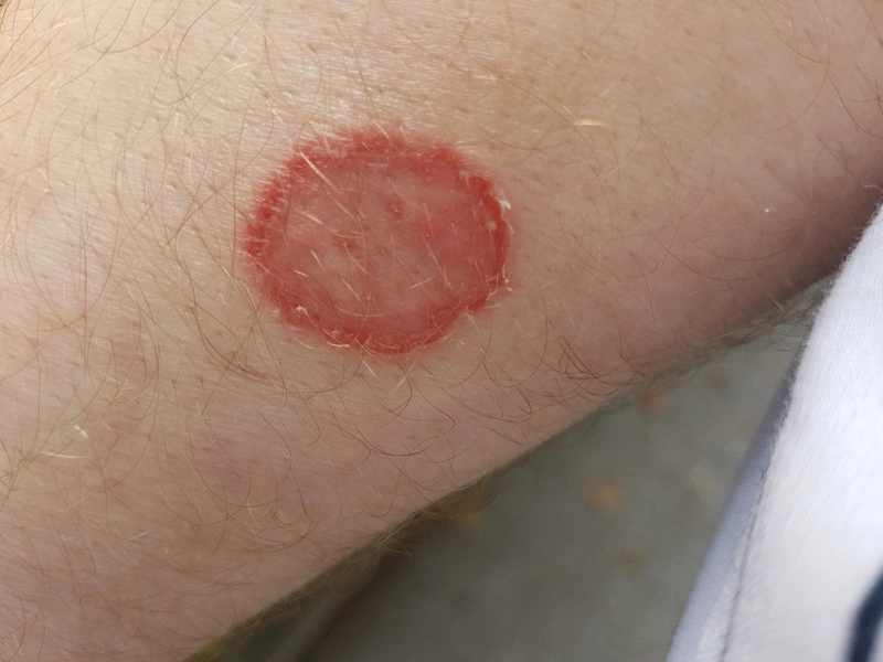
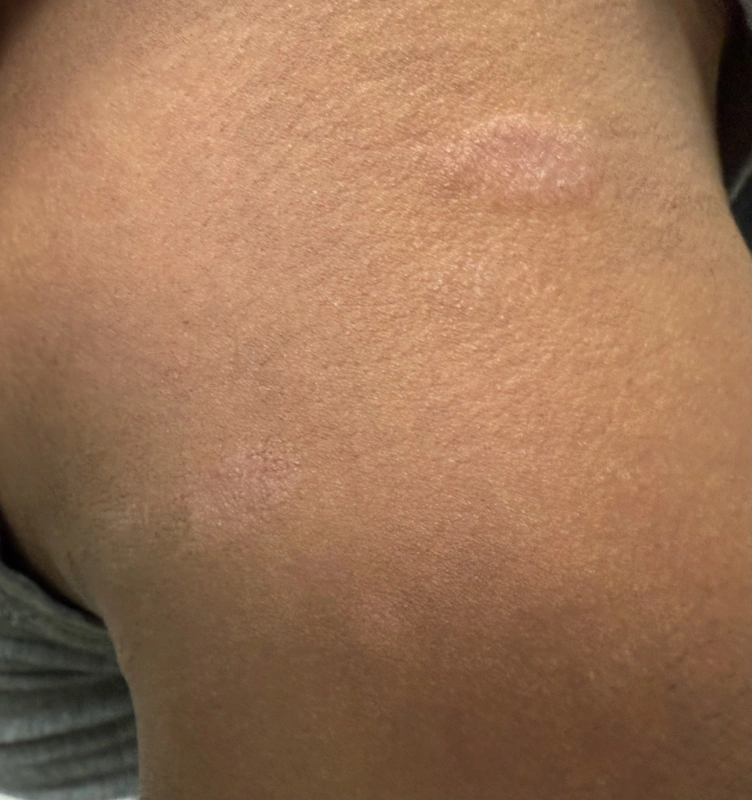
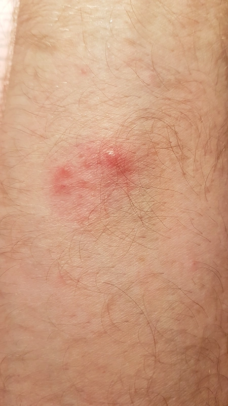
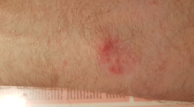
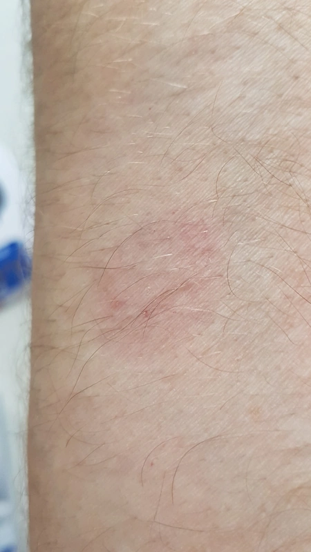
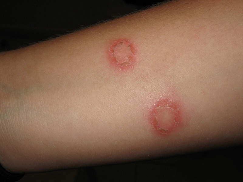
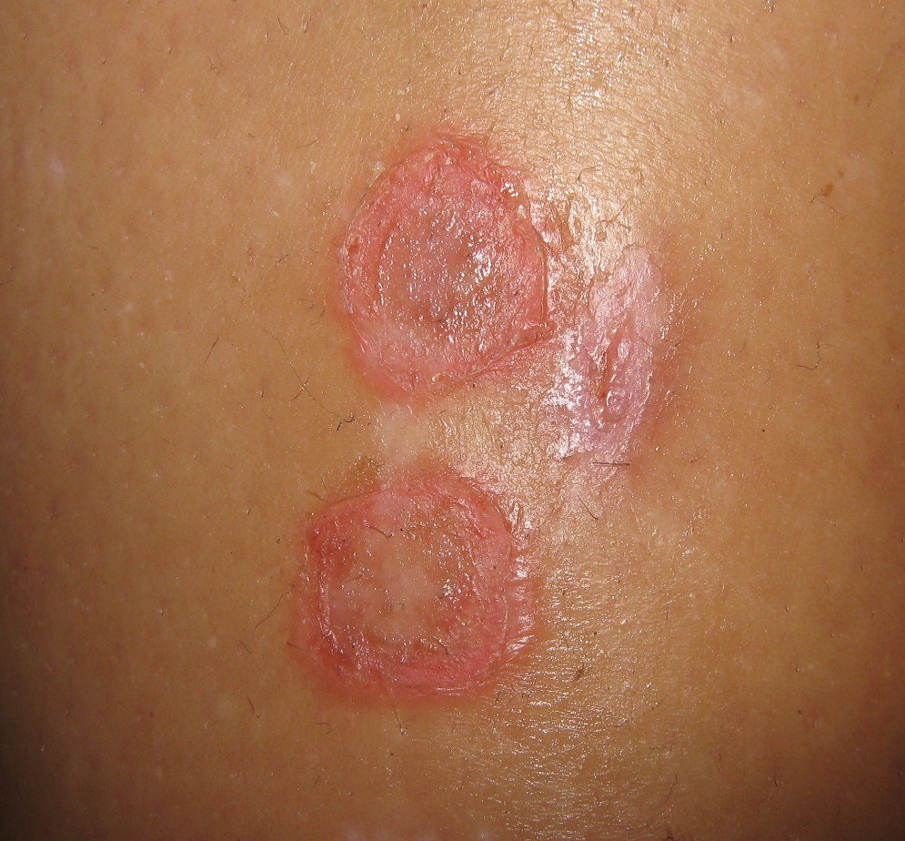
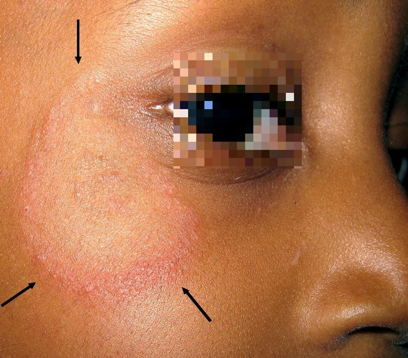
Definition
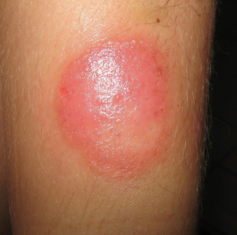
Tinea corporis is a dermatophyte infection of the glabrous skin of the trunk and limbs (excluding face, hands, feet and groin). Because of its annular lesions it is also called "ringworm".
- Clinical features: annular sharply demarcated erythematosquamous plaques with active border and central clearing, often pruritus
- Pathogens: mainly T. rubrum, also T. interdigitale/mentagrophytes, T. tonsurans, M. canis
Occurrence & epidemiology
It is transmitted from person to person (anthropophilic species such as Trichophyton rubrum and T. tonsurans, e.g. in contact sports), from animals (zoophilic species such as Microsporum canis from cats and dogs, less often T. verrucosum from cattle or T. erinacei from hedgehogs) and via shared towels, bedding and clothing or by spread from tinea pedis. Zoophilic species can cause highly inflammatory lesions (kerion). Risk factors include diabetes mellitus, immunodeficiency (disseminated disease in HIV), hyperhidrosis and pets.
Diagnosis
- Direct microscopy with potassium hydroxide (KOH) from the scaly active border, fungal culture to identify the organism (pointing to the source of infection); examination of the whole skin including scalp and nails.
- Distinction from discoid eczema, psoriasis, the herald patch of pityriasis rosea and seborrhoeic dermatitis; an atypical, ill-defined picture without scale and erythema as "tinea incognito".
Differential diagnoses
 Nummular eczema (discoid eczema)
 Atopic dermatitis (atopic eczema)
 Psoriasis vulgaris (plaque psoriasis)
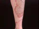 Granuloma annulare
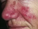 Rosacea
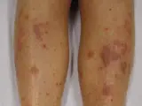 Lichen planus
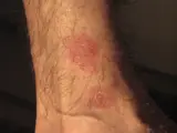 Pityriasis rosea
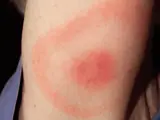 Erythema migrans (Lyme disease)
Image credits for the thumbnails
- Nummular eczema (discoid eczema): Ifnord, “Nummular dermatitits”, CC BY-SA 4.0, via Wikimedia Commons · CC BY-SA 4.0 · Source
- Atopic dermatitis (atopic eczema): Eisfelder, “Atopic dermatitis child”, CC BY-SA 3.0, via Wikimedia Commons · CC BY-SA 3.0 · Source
- Psoriasis vulgaris (plaque psoriasis): MediaJet, “An Arm Covered With Plaque Type Psoriasis”, CC BY-SA 3.0, via Wikimedia Commons · CC BY-SA 3.0 · Source
- Granuloma annulare: Kreuter et al., “Generalized granuloma annulare”, CC BY-SA 2.0, via Wikimedia Commons · CC BY-SA 2.0 · Source
- Rosacea: Michael Sand, Daniel Sand, Christina Thrandorf, Volker Paech, Peter Altmeyer, Falk G Bechara, “Rosacea”, CC BY 2.5, via Wikimedia Commons, cropped · CC BY 2.5 · Source
- Lichen planus: James Heilman, MD, “Lichen Planus (2)”, CC BY-SA 3.0, via Wikimedia Commons · CC BY-SA 3.0 · Source
- Pityriasis rosea: Aceofhearts1968, “Pityriasis rosea-1”, Public Domain, via Wikimedia Commons · Public domain · Source
- Erythema migrans (Lyme disease): James Gathany (CDC), “Erythema migrans - erythematous rash in Lyme disease - PHIL 9875”, Public Domain, via Wikimedia Commons · Public domain · Source · CDC PHIL #9875
Treatment principles
- Localised lesions are treated with topical antifungals, extending beyond the visible margins and continued for some time after healing.
- Extensive disease, follicular involvement, immunosuppression or treatment failure require systemic therapy after identification of the organism.
- Source animals are treated; topical glucocorticoids alone should be avoided as they mask the clinical picture (tinea incognito).
- In extensive or treatment-resistant tinea, resistant organisms such as Trichophyton indotineae should be considered; culture and susceptibility testing are then particularly important.
Specific treatment regimens with drugs and doses are covered in the DermaFuchs app.
Keep learning in the app
References (selection)
Cross-references
Mentioned in
Keep learning: test yourself
One question from the DermaFuchs app on Tinea corporis – tap an answer.
Question 1 of 1
Which activity is a known risk factor for Tinea corporis, alongside animal contact and shaving trauma?
Answer
Correct: contact sports. Contact sports (like wrestling) facilitate direct skin-to-skin transmission of dermatophytes (e.g., Tinea gladiatorum).
More questions like these – with explanations and spaced repetition – in the app.
Note: This page is intended for medical education and does not replace diagnosis or treatment decisions in individual cases.