Nummular eczema (discoid eczema)
- Synonyms
- Münzekzem, diskoides Ekzem, Ekzema nummulare, mikrobielles Ekzem, nummular eczema, discoid eczema
- Specialty
- Dermatology · Allergies
- Images
- Clinical 4
- In the app
- 1 flashcard
- Last updated
- 10/2026 · Dr. Pascal Bafteh
Contents
Images (4)
Definition
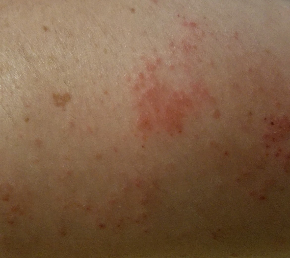
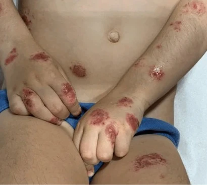
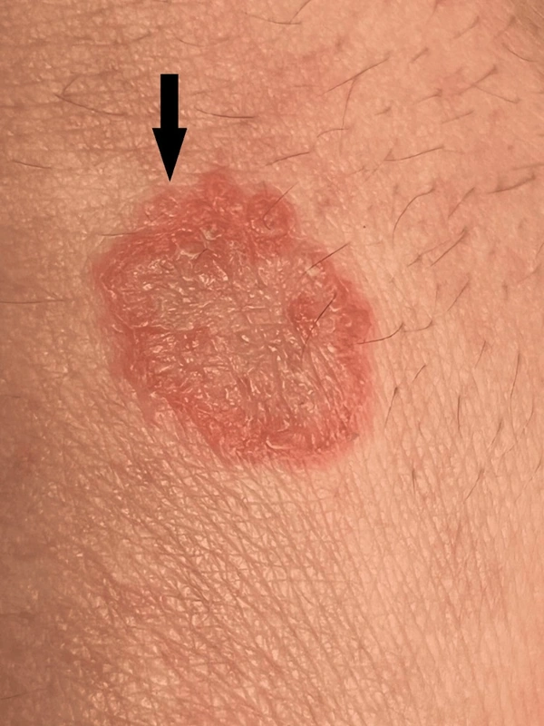
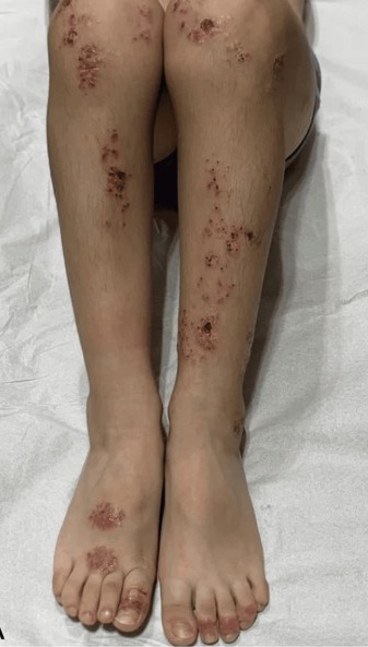
Nummular (discoid) eczema is a chronic relapsing form of eczema with sharply demarcated, round, coin-sized plaques. The cause is unknown; some cases are associated with Staphylococcus aureus. Triggers include local injury (scratch, insect bite, burn), impetigo or wound infection, contact dermatitis, dry skin and varicose veins; associations exist with atopic eczema and eczema craquelé.
Occurrence & epidemiology
It occurs at all ages, slightly more often in older men and younger women; in men there is an association with chronic alcohol use. Drugs that dry the skin can trigger it; relapses occur especially in the cold winter months.
Clinical features
- Round to oval, mostly 1–3 cm, well-circumscribed, inflamed plaques: acute exudative with oozing papules and blisters, subacute to chronic as dry red plaques; the skin between the patches is usually dry.
- Mainly on the limbs, especially the legs, often bilateral, but may be widespread; usually very itchy; in severe cases generalisation through autoeczematisation.
- Chronic course with frequent relapses.
Diagnosis
- Clinical diagnosis.
- Distinction from tinea corporis (mycology), plaque psoriasis and pityriasis rosea; bacterial swab (S. aureus); patch testing in chronic disease (contact allergy to nickel and chromate commonly reported).
Differential diagnoses
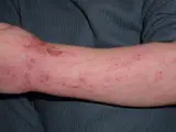 Atopic dermatitis (atopic eczema)
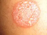 Tinea corporis
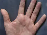 Dyshidrotic eczema (pompholyx)
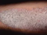 Psoriasis vulgaris (plaque psoriasis)
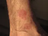 Pityriasis rosea
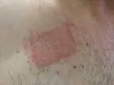 Contact dermatitis (contact eczema)
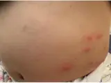 Papular urticaria
Image credits for the thumbnails
- Atopic dermatitis (atopic eczema): Eisfelder, “Atopic dermatitis child”, CC BY-SA 3.0, via Wikimedia Commons · CC BY-SA 3.0 · Source
- Tinea corporis: Corina G., “Tinea corporis”, Public Domain, via Wikimedia Commons · Public domain · Source
- Dyshidrotic eczema (pompholyx): Inwe (Wikimedia Commons), “Dyshidrosis”, CC BY-SA 3.0, via Wikimedia Commons · CC BY-SA 3.0 · Source
- Psoriasis vulgaris (plaque psoriasis): MediaJet, “An Arm Covered With Plaque Type Psoriasis”, CC BY-SA 3.0, via Wikimedia Commons · CC BY-SA 3.0 · Source
- Pityriasis rosea: Aceofhearts1968, “Pityriasis rosea-1”, Public Domain, via Wikimedia Commons · Public domain · Source
- Contact dermatitis (contact eczema): Dr.khatmando, “Severe contact dermatitis buprenorphine transdermal patch”, CC BY-SA 4.0, via Wikimedia Commons · CC BY-SA 4.0 · Source
- Papular urticaria: Sekerel BE, Ilgun Gurel D, Sahiner UM et al. (Front Allergy 2023), CC BY 4.0, via PubMed Central (PMC10655015) · CC BY 4.0 · Source
Treatment principles
- Nummular eczema is often persistent; the basis is consistent emollient care and avoidance of irritants.
- First-line treatment is moderate- to high-potency topical glucocorticoids, alternatively topical calcineurin inhibitors, with antiseptics or antibiotics added for superinfection.
- Extensive or refractory disease may be treated with phototherapy or short-term systemic therapy; contact allergy should be excluded where appropriate.
Specific treatment regimens with drugs and doses are covered in the DermaFuchs app.
Keep learning in the app
References (selection)
Cross-references
Mentioned in
- Anal eczema
- Atopic dermatitis (atopic eczema)
- Granuloma annulare
- Necrolytic migratory erythema
- Guttate psoriasis
- Psoriasis vulgaris (plaque psoriasis)
- Caterpillar dermatitis caused by oak processionary moth (Thaumetopoea processionea)
- Tinea corporis
- X-linked recessive ichthyosis
- Prurigo nodularis
- Insect bite and sting reaction (local and large local reaction)
Keep learning
Keep learning with flashcards, exam questions and spot diagnoses in the DermaFuchs app – free, in your browser or as an app.
Related pages: Atopic dermatitis (atopic eczema) · Tinea corporis · Dyshidrotic eczema (pompholyx)
Note: This page is intended for medical education and does not replace diagnosis or treatment decisions in individual cases.