Mastocytosis (urticaria pigmentosa)
Exam relevance: in 7 of more than 50 board exam reports · rank 19
- Synonyms
- Urticaria pigmentosa, mastocytosis
- Specialty
- Dermatology · Allergies
- Images
- Clinical 6 · Dermoscopy 2 · Histology 1
- In the app
- 8 flashcards
- Last updated
- 10/2026 · Dr. Pascal Bafteh
Contents
Images (9)
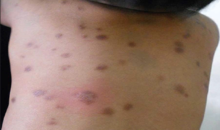
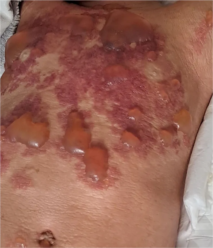
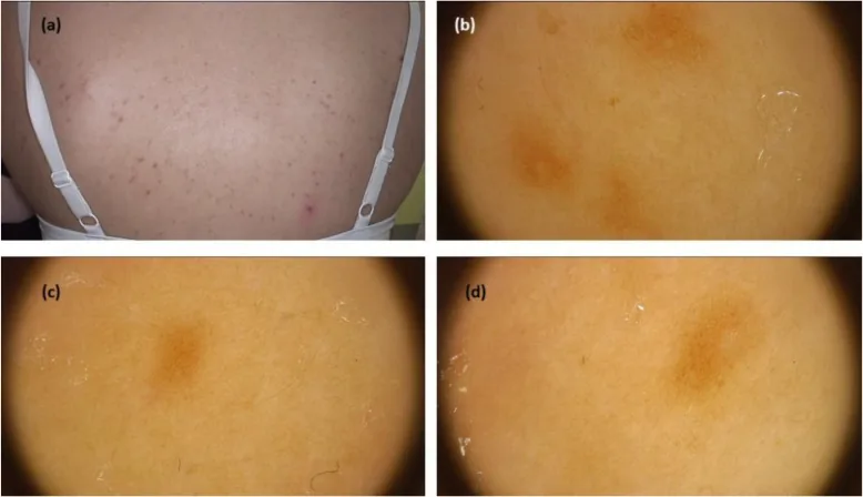 Dermoscopy
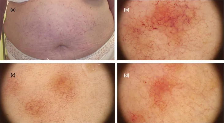 Dermoscopy
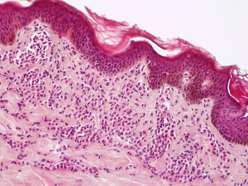 Histology
Definition
In mastocytosis, mast cells multiply and accumulate in the skin, the bone marrow and sometimes other organs. Mast cells store mediators such as histamine and tryptase; when these are released, itch, wheals, flushing, abdominal symptoms and, in the extreme, circulatory shock occur. In adults an acquired change in the KIT gene is almost always the cause, mostly the point mutation D816V; in children it is found in about three out of four cases. The disease is not contagious and in the vast majority of cases not malignant.
Types
The WHO classification (5th edition, 2022) distinguishes three groups:
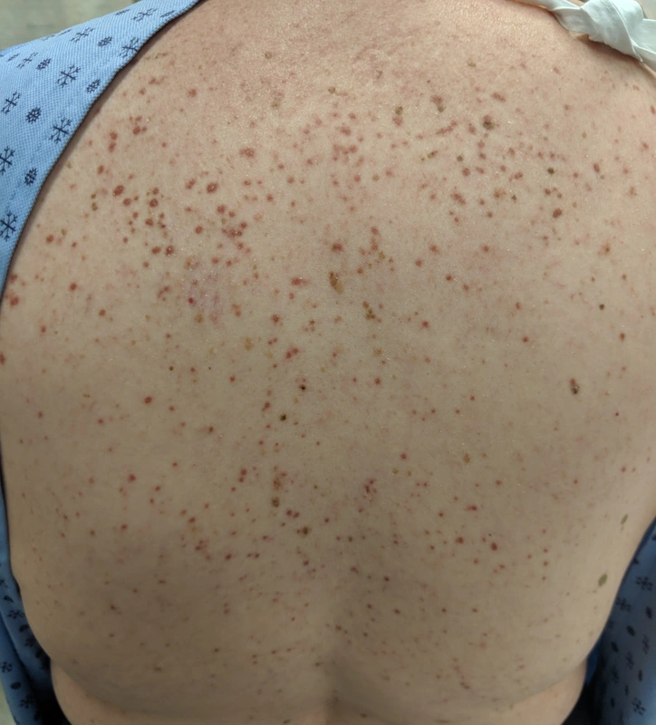
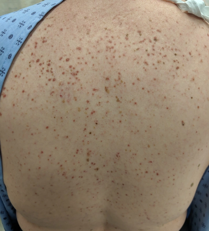
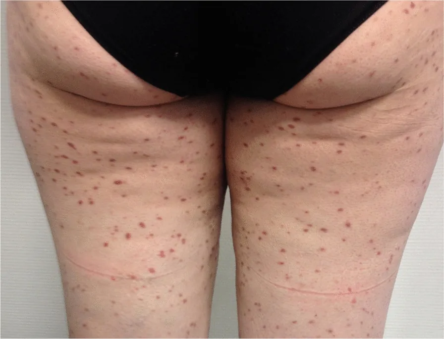
- Cutaneous mastocytosis – only the skin is affected, typical of children. It has three forms: maculopapular cutaneous mastocytosis (formerly urticaria pigmentosa) with many brown spots or papules, in children often larger and of varying size (polymorphic), in adults small and uniform (monomorphic); the rare diffuse cutaneous mastocytosis of infancy, in which the whole skin is thickened, doughy and often blistering; and mastocytoma, a single (up to three) yellowish-brown nodule or plaque.
- Systemic mastocytosis – mast cells also in the bone marrow or other organs, typical of adults. The most common form is indolent systemic mastocytosis; since 2022 the WHO lists the form confined to the bone marrow separately; less common are smouldering and aggressive forms, the form with an associated haematological neoplasm and mast cell leukaemia.
- Mast cell sarcoma – an extremely rare malignant tumour.
Occurrence
Mastocytosis is rare; estimates are around one in 10,000 people. In most affected children the skin lesions appear in the first two years of life, and in many they regress markedly or disappear by puberty. If mastocytosis starts in adulthood, however, it usually persists, and bone marrow involvement is then almost always found.
Clinical features
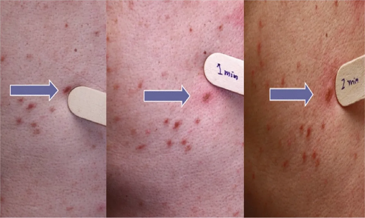
Skin: reddish-brown to yellowish macules, papules or plaques, mainly on the trunk and thighs; face, palms and soles are usually spared. Rubbing a lesion makes it swell within minutes into an itchy wheal with a red flare (Darier's sign). In infants, blisters can form within the lesions.
Mediator symptoms: itch, attacks of redness of face and upper body (flushing), abdominal cramps, diarrhoea, nausea, headache, fatigue and palpitations. Common triggers are heat, friction, exercise, alcohol, certain painkillers, anaesthesia and radiocontrast media.
Anaphylaxis: adults with systemic mastocytosis in particular have an increased risk of severe allergic reactions, above all after wasp or bee stings. Conversely, a severe sting reaction with circulatory involvement can be the first sign of a previously unknown mastocytosis.
Other organs: in the systemic form, osteoporosis and bone pain are common; liver, spleen and lymph nodes can be enlarged.
Diagnosis
- Skin: clinical picture plus Darier's sign; confirmation by skin biopsy showing increased mast cells (stains for tryptase and CD117). Rubbing the lesion before the biopsy empties the mast cells and can blur the findings.
- Serum tryptase (baseline): usually normal in children with purely cutaneous disease; a persistently raised value above 20 ng/ml in adults suggests systemic mastocytosis. Hereditary alpha-tryptasaemia can also raise the value.
- KIT D816V: highly sensitive testing in blood, often positive in adults.
- Bone marrow biopsy: in adults with skin involvement, with tryptase above 20 ng/ml or after unexplained anaphylaxis. The WHO criteria require dense mast cell aggregates in the bone marrow (major criterion) and/or minor criteria such as atypical, spindle-shaped mast cells, expression of CD25, CD2 or CD30, the KIT mutation and raised tryptase.
- In systemic disease: bone density measurement, blood count, liver tests, abdominal ultrasound.
In children with a typical skin form and normal tryptase, a bone marrow examination is usually not needed.
Differential diagnosis
- Café-au-lait macules, lentigines, melanocytic naevi: brownish spots, but no Darier's sign.
- Urticaria: wheals disappear within hours without brownish residual spots.
- Juvenile xanthogranuloma: yellow-orange papules in toddlers, often on the head.
- Insect bite reactions, prurigo: itchy papules with scratch marks, changing location.
- Blisters in infants: bullous impetigo, staphylococcal scalded skin syndrome, epidermolysis bullosa.
- Flushing: carcinoid syndrome, rosacea, menopause, drugs.
Differential diagnoses
 Urticaria
 Atopic dermatitis (atopic eczema)
 Pityriasis rosea
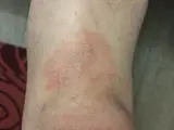 Mycosis fungoides
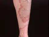 Granuloma annulare
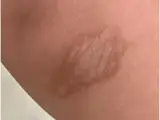 Mastocytoma
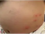 Papular urticaria
- Giant urticaria
Image credits for the thumbnails
- Urticaria: Gzzz, “Urticaria child”, CC BY-SA 4.0, via Wikimedia Commons · CC BY-SA 4.0 · Source
- Atopic dermatitis (atopic eczema): Eisfelder, “Atopic dermatitis child”, CC BY-SA 3.0, via Wikimedia Commons · CC BY-SA 3.0 · Source
- Pityriasis rosea: Aceofhearts1968, “Pityriasis rosea-1”, Public Domain, via Wikimedia Commons · Public domain · Source
- Mycosis fungoides: Wikindrea, “Mycosis Fungoides Before Sun and Cartamo”, CC BY-SA 4.0, via Wikimedia Commons · CC BY-SA 4.0 · Source
- Granuloma annulare: Kreuter et al., “Generalized granuloma annulare”, CC BY-SA 2.0, via Wikimedia Commons · CC BY-SA 2.0 · Source
- Mastocytoma: Altaweel F, Mosbeh A, Alenezi H et al. (JAAD Case Rep 2026), CC BY 4.0, via PubMed Central (PMC13396385) · CC BY 4.0 · Source
- Papular urticaria: Sekerel BE, Ilgun Gurel D, Sahiner UM et al. (Front Allergy 2023), CC BY 4.0, via PubMed Central (PMC10655015) · CC BY 4.0 · Source
Treatment principles
- The basis is avoidance of triggers of mast cell degranulation; in systemic mastocytosis or with a history of anaphylaxis, an emergency kit with an auto-injector is mandatory, and in hymenoptera venom allergy lifelong venom immunotherapy is additionally recommended.
- Symptomatic treatment comprises H1 and H2 antihistamines, mast cell stabilisers and leukotriene receptor antagonists, with topical or intralesional glucocorticoids for skin lesions.
- Advanced systemic forms are treated by haematologists with targeted KIT tyrosine kinase inhibitors, and in selected cases with allogeneic stem cell transplantation; KIT tyrosine kinase inhibitors are now also approved for indolent systemic forms when moderate to severe symptoms persist despite symptomatic treatment.
Specific treatment regimens with drugs and doses are covered in the DermaFuchs app.
Keep learning in the app
References (selection)
- Hartmann K, Escribano L, Grattan C et al. Cutaneous manifestations in patients with mastocytosis: consensus report of the European Competence Network on Mastocytosis, the American Academy of Allergy, Asthma & Immunology, and the European Academy of Allergology and Clinical Immunology. J Allergy Clin Immunol. 2016;137(1):35-45
- Khoury JD, Solary E, Abla O et al. The 5th edition of the World Health Organization Classification of Haematolymphoid Tumours: Myeloid and Histiocytic/Dendritic Neoplasms. Leukemia. 2022;36(7):1703-1719
- Pardanani A. Systemic mastocytosis in adults: 2023 update on diagnosis, risk stratification and management. Am J Hematol. 2023;98(7):1097-1116
Cross-references
Keep learning: test yourself
Two questions from the DermaFuchs app on Mastocytosis (urticaria pigmentosa) – tap an answer.
Question 1 of 2
Which mutation is predominantly found in adults with systemic mastocytosis?
Answer
Correct: KIT-D816V mutation. The KIT-D816V mutation is predominantly found in adults with systemic mastocytosis, affecting bone marrow and organs.
Question 2 of 2
What are the characteristic clinical findings of Urticaria pigmentosa on the trunk and proximal extremities?
Answer
Correct: brown macules + Darier's sign. Urticaria pigmentosa classically presents as brown macules that exhibit Darier's sign (urtication upon rubbing) due to mast cell degranulation.
More questions like these – with explanations and spaced repetition – in the app.
Note: This page is intended for medical education and does not replace diagnosis or treatment decisions in individual cases.