Erysipeloid
- Synonyms
- fish handler disease, Erysipelothrix infection
- Specialty
- Dermatology · Bacterial infections
- Images
- Clinical 2
- In the app
- 2 flashcards
- Last updated
- 10/2026 · Dr. Pascal Bafteh
Contents
Images (2)
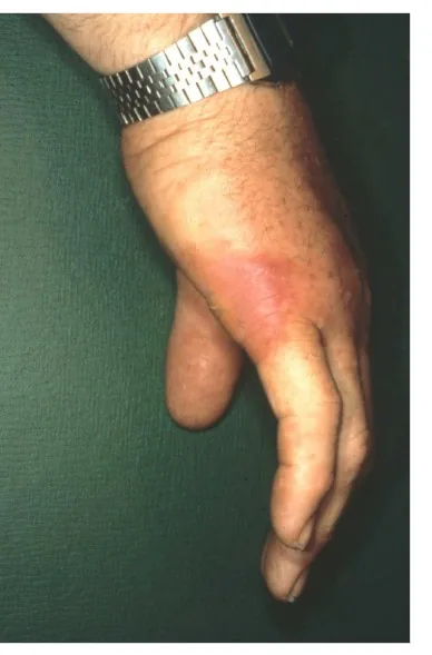
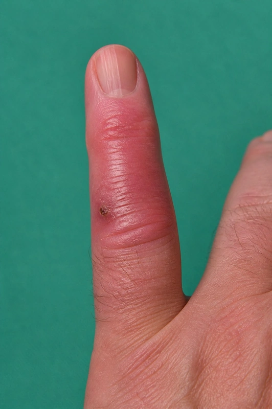 AI illustration
Definition
Typical is a livid-red sharply demarcated lesion on hands / fingers (swine erysipelas, zoonosis caused by Erysipelothrix rhusiopathiae, gram-positive rod).
Risk group are fishermen + butchers — often after skin injury when handling raw meat/fish.
Clinical features
- A sharply demarcated, violaceous red, raised, shiny erythema develops at the entry site (usually finger or back of the hand), slowly spreading outwards over a few days.
- Burning, itching or pain; regional lymphadenopathy and lymphangitis in about one third, mild fever only in a few. Rarely systemic courses, usually as septic arthritis or endocarditis (often of the aortic valve).
Diagnosis
- Occupational and exposure history (meat, fish, shellfish, animal contact) and clinical picture.
- Pathogen detection by culture of a full-thickness biopsy (the organisms lie in the deeper skin layers) or PCR; blood cultures if endocarditis is suspected, though they are rarely positive.
Differential diagnoses
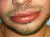
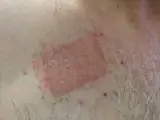
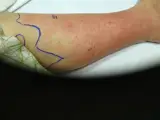
Image credits for the thumbnails
- Erysipelas: Poupou l'quourouce, “Erysipel”, CC BY-SA 3.0, via Wikimedia Commons · CC BY-SA 3.0 · Source
- Urticaria: Gzzz, “Urticaria child”, CC BY-SA 4.0, via Wikimedia Commons · CC BY-SA 4.0 · Source
- Angioedema: James Heilman, MD, “AngioedemaFra”, CC BY-SA 3.0, via Wikimedia Commons · CC BY-SA 3.0 · Source
- Contact dermatitis (contact eczema): Dr.khatmando, “Severe contact dermatitis buprenorphine transdermal patch”, CC BY-SA 4.0, via Wikimedia Commons · CC BY-SA 4.0 · Source
- Cellulitis (phlegmon): Stephen Ewen, “Left cellulitis of leg”, CC BY-SA 4.0, via Wikimedia Commons · CC BY-SA 4.0 · Source
Treatment principles
- The causative organism, Erysipelothrix rhusiopathiae, is highly susceptible to antibiotics; treatment is systemic, usually with penicillins.
- Although the localised cutaneous form can resolve spontaneously, treatment shortens the course and prevents rare systemic complications such as endocarditis.
- Rest and elevation relieve symptoms.
Specific treatment regimens with drugs and doses are covered in the DermaFuchs app.
Keep learning in the app
References (selection)
Cross-references
Keep learning
Keep learning with flashcards, exam questions and spot diagnoses in the DermaFuchs app – free, in your browser or as an app.
Related pages: Erysipelas · Urticaria · Angioedema
Note: This page is intended for medical education and does not replace diagnosis or treatment decisions in individual cases.