Urticaria
Definition & Classification
- Acute Urticaria: < 6 weeks. Chronic Spontaneous Urticaria (CSU): ≥ 6 weeks.
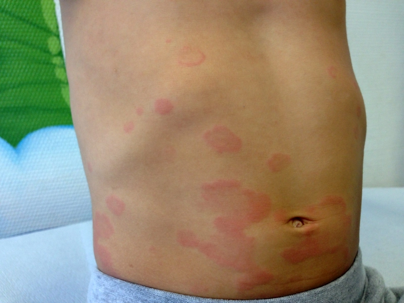
- Wheals: transient (< 24 hours), itchy, due to mast cell degranulation.
Pathomechanism
The common final pathway
- One cell decides: Whatever the initial trigger, the end point is always degranulation of the cutaneous mast cell. That is why the weal looks the same in every form, and why the same treatment works across very different causes.
- What is released: immediately from preformed granules histamine, tryptase and heparin; newly synthesised within minutes prostaglandin D2, leukotrienes C4/D4 and platelet-activating factor; over hours cytokines (TNF-α, IL-4, IL-5, IL-13).
- What histamine does in the skin: Via the H1 receptor on endothelium and sensory C fibres it produces vasodilation (redness), increased vascular permeability (weal) and stimulation of itch fibres (pruritus) — the three components of Lewis's triple response.
- Why the weal is fleeting: it is pure oedema of the upper dermis without cell death; once the fluid has drained it disappears without trace — hence the rule that an individual weal lasts under 24 hours. A lesion that persists longer or leaves pigment is not ordinary urticaria but raises the suspicion of urticarial vasculitis — the livid, brownish-violet hue comes from haemosiderin after red cell extravasation and is exactly what the fleeting weal lacks.
- Angio-oedema as the sibling finding: the same mediator release deeper down, in the subcutis and submucosa — hence tightness rather than itch there, and a distinctly slower resolution (up to 72 hours).
How the mast cell is switched on — two endotypes of CSU
In chronic spontaneous urticaria two routes are now distinguished by which the body activates its own mast cells. Both end at the same cell but differ in the antibody, the target antigen and the response to treatment.
- Type I — "autoallergic" (IgE against self): The body produces IgE antibodies against its own structures ("autoallergens", e.g. thyroid peroxidase, IL-24, tissue factor). This IgE sits on FcεRI just like ordinary allergen-specific IgE — only the "allergen" is one of the body's own molecules. The cell is therefore triggered through the entirely normal type I route, without there ever having been contact with an external allergen.
- Type IIb — "autoimmune" (IgG against the receptor): Here IgG autoantibodies bind directly to FcεRI or to the IgE sitting on it and cross-link neighbouring receptors. The cross-linking triggers degranulation; activated complement (C5a) amplifies the effect.
- The decisive difference from type IIa: Classic type II (IIa) destroys the target cell (cytolysis, as in immune thrombocytopenia or autoimmune haemolytic anaemia). Type IIb by contrast only alters the function of the target cell — it is switched on, not killed. The same logic applies in Graves disease, where the autoantibody stimulates rather than destroys the TSH receptor.
Why the distinction matters in practice
- Diagnosis: Features pointing to type IIb are a low total IgE, positive anti-TPO IgG, basopenia and a positive autologous serum test or basophil activation test.
- What the model explains: the association of CSU with autoimmune thyroiditis, the female predominance, and the observation that infections, stress and NSAIDs are not causes but amplifiers of an already primed mast cell. NSAIDs act not allergically but by shunting arachidonic acid metabolism towards the leukotrienes (pseudoallergic).
The classic classification in brief
- Type I (allergic): IgE-mediated via FcεRI on mast cells — the rule in acute urticaria (food, drugs, insect venom).
- Autoimmune (type IIb): autoantibodies against IgE or FcεRI. Diagnosis: autologous serum skin test (ASST).
- Mediator: histamine → vasodilation, oedema, pruritus.
Differential diagnoses
- Angioedema
- Fixed drug eruption (FDE)
- Urticarial vasculitis
- Urticaria papulosa
- Atopic dermatitis (atopic eczema)
- Allergic contact dermatitis
- Giant urticaria
Practise Urticaria in the app
Flashcards with spaced repetition, exam questions and spot-the-diagnosis on this topic – in DermaFuchs, free of charge.
In the DermaFuchs app: 2 flashcards · 7 clinical images
Open in browser Download on the App StoreSources (selection)
- Kaplan et al. 2023
- Kolkhir P, Bonnekoh H, Metz M, Maurer M. Chronic Spontaneous Urticaria: A Review. JAMA. 2024;332(17):1464-1477.
- AWMF S3 Urtikaria 2022
More allergies and eczemas
- Angioedema
- Atopic dermatitis (atopic eczema)
- Mastocytosis (urticaria pigmentosa)
- Seborrhoeic dermatitis
Note: This page is intended for medical education and does not replace diagnosis or treatment decisions in individual cases. Treatment and follow-up content is available in the app.