Dysplastic naevus (atypical naevus)
- Synonyms
- atypischer Nävus, Clark-Nävus, auffälliges Muttermal, Syndrom der dysplastischen Nävi, FAMMM, atypical mole, dysplastic nevus, atypical naevus, Clark naevus
- Specialty
- Dermatology · Skin tumours
- Images
- Clinical 4 · Histology 1
- Last updated
- 10/2026 · Dr. Pascal Bafteh
Contents
Images (5)
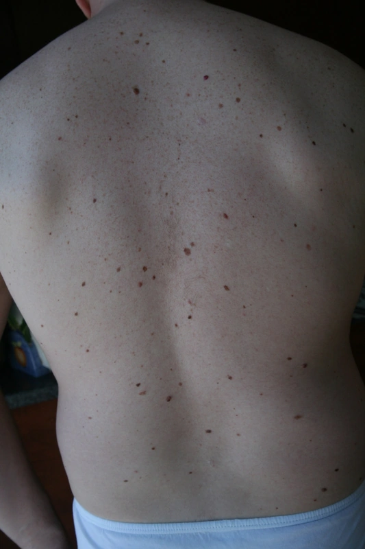
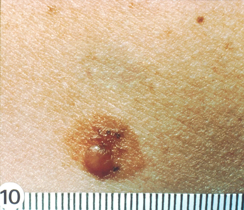
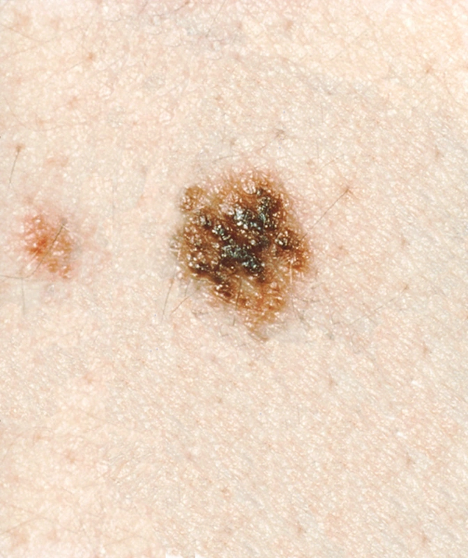
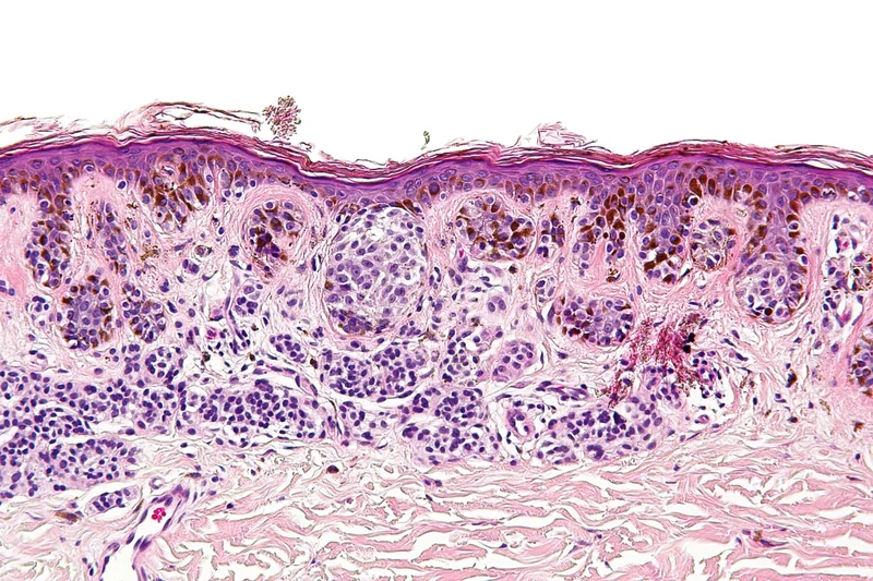 Histology
Definition
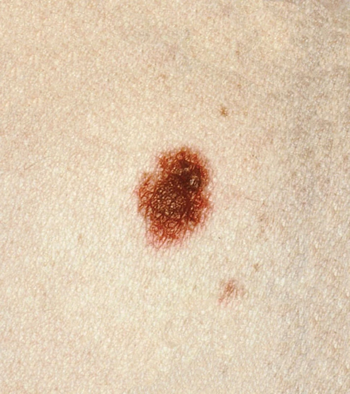
Atypical (clinically irregular) naevi are acquired, benign melanocytic naevi with unusual features such as asymmetry, irregular borders, colour variation or a diameter over 5 mm. "Dysplastic naevus", by contrast, denotes a histological finding (architectural disorder and cytological atypia of the melanocytes); the two terms are often used interchangeably but are not identical.
Occurrence & epidemiology
Atypical naevi are common (prevalence about 5–12% in white populations) and usually develop in childhood and early adolescence. Multiple atypical naevi – especially as part of the dysplastic naevus syndrome (familial atypical multiple mole melanoma syndrome, FAMMM) with germline mutations, e.g. in CDKN2A – are associated with a markedly increased melanoma risk. A single dysplastic naevus, by contrast, rarely progresses to melanoma; it is mainly a risk marker.
Clinical features
- Usually over 5 mm, with ill-defined or irregular borders, asymmetrical, with variegated colours; predominantly flat, a central papule may give a "fried-egg" appearance.
Diagnosis
- Dermoscopy with pattern comparison; digital documentation for comparison over time.
- An outlier naevus ("ugly duckling") or eccentric peripheral hypo-/hyperpigmentation calls for histological examination to rule out melanoma; the diagnosis of "dysplastic naevus" is histological.
- Family history of melanoma and total naevus count to assess risk.
Differential diagnoses
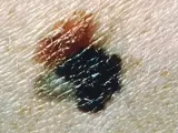 Melanoma
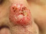 Basal cell carcinoma
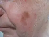 Lentigo maligna
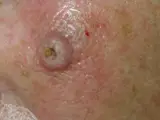 Keratoacanthoma
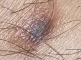 Dermatofibroma
 Blue naevus
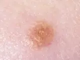 Spitz naevus (spindle and epithelioid cell naevus)
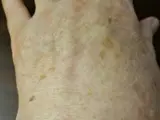 Solar lentigo (age spot)
Image credits for the thumbnails
- Melanoma: National Cancer Institute (unknown photographer), “Melanoma (4)”, Public Domain, via Wikimedia Commons · Public domain · Source
- Basal cell carcinoma: Bin im Garten, “Basaliom am Nasenrücken 2”, CC BY-SA 3.0, via Wikimedia Commons · CC BY-SA 3.0 · Source
- Lentigo maligna: Omar Bari, Philip R. Cohen, “Photograph of lentigo maligna melanoma”, CC BY 3.0, via Wikimedia Commons · CC BY 3.0 · Source
- Keratoacanthoma: Klaus D. Peter, Wiehl, Germany, “Spinaliom”, CC BY 3.0 DE, via Wikimedia Commons · CC BY 3.0 DE · Source
- Dermatofibroma: Jmarchn, “Pigmented and tenderness dermatofibroma”, CC BY-SA 3.0, via Wikimedia Commons · CC BY-SA 3.0 · Source
- Blue naevus: Dannii Brighton, “Blue Nevus”, CC BY-SA 3.0, via Wikimedia Commons · CC BY-SA 3.0 · Source
- Spitz naevus (spindle and epithelioid cell naevus): The Armed Forces Institute of Pathology (AFIP), Public domain, via Wikimedia Commons · Public domain · Source
- Solar lentigo (age spot): Beyond My Ken, “Liver spots, light-skinned Caucasian man, age 63”, CC BY-SA 4.0, via Wikimedia Commons · CC BY-SA 4.0 · Source
Treatment principles
- Prophylactic removal of all dysplastic naevi is not appropriate.
- Regular dermatological examinations with dermoscopy are recommended, supplemented by total body photography or digital monitoring in patients with many naevi, together with consistent photoprotection.
- Lesions showing clinically or dermoscopically suspicious changes are excised completely and examined histologically.
Specific treatment regimens with drugs and doses are covered in the DermaFuchs app.
Keep learning in the app
References (selection)
Cross-references
Keep learning
Keep learning with flashcards, exam questions and spot diagnoses in the DermaFuchs app – free, in your browser or as an app.
Related pages: Melanoma · Basal cell carcinoma · Lentigo maligna
Note: This page is intended for medical education and does not replace diagnosis or treatment decisions in individual cases.