Dermatofibrosarcoma protuberans
Definition & Epidemiology
- Most common sarcoma of the skin, fibroblastic differentiation with (myo-)fibroblastic components.
- Average age: ~ 40 years, slow destructive local growth.
- Metastasis rare: < 5 % hematogenous pulmonary.
Genetics & Pathogenesis
- Characteristic translocation: [t COL1A1-PDGFB|t EWSR1-FLI1|BRAF-V600E|MYCN amplification] in > 90 % → constitutive activation of the PDGF-β receptor signaling pathway → molecular target forImatinib.
- Sporadic, no known heritability.
Clinical Presentation
- Location: trunk-predominant (~ 50 %), proximal extremities, shoulders; rarely head and neck.
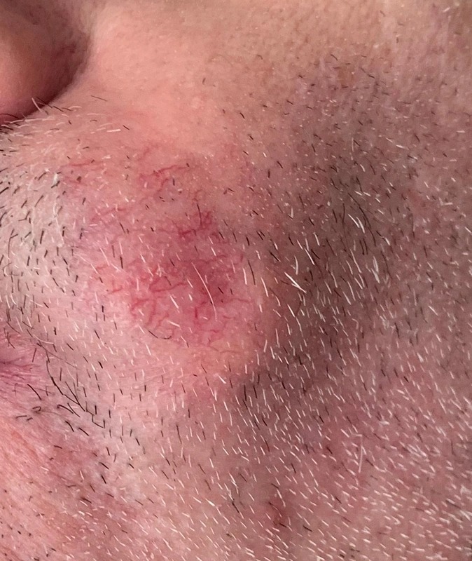
- Appearance: brownish-livid plaque.
- Course: Years of unremarkable plaque-stage progression until nodular manifestation; destructive-infiltrative, R0 resection technically challenging due to tentacular subcutaneous extensions.
Sub-Variants
- Classic DFSP — as above.
- Bednar tumor (pigmented DFSP variant): melanin-containing dendritic cells.
- Fibrosarcomatous DFSP transformation (DFSP-FS): Dedifferentiation + Mitosis ↑.
- Giant cell fibroblastoma: pediatric variant, same COL1A1-PDGFB translocation.
Diagnostics
- Clinical presentation + ultrasound of the primary tumor (depth of extension, hypoechoic-heterogeneous).
- MRI in case of suspected deep infiltration (subcutis, fascia).
- Punch or incisional biopsy: Histology shows monomorphic spindle cell proliferation in a storiform pattern, subcutaneous infiltration as a "honeycomb pattern".
- Immunohistochemistry: CD34+ + CD99+, F XIIIa− for differentiation from dermatofibroma (= XIIIa+).
- Molecular: FISH or RT-PCR for COL1A1-PDGFB fusion to confirm difficult cases (e.g., fibrosarcomatous variant).
- Staging: Whole-body examination + ultrasound of regional lymph nodes; for DFSP-FS, additionally CT thorax (metastases rare, but mandatory).
Differential diagnoses
- Dermatofibroma
- Lipoma
- Epidermal cyst (atheroma)
- Basal cell carcinoma
- Melanoma
- Cutaneous angiosarcoma
- Kaposi's sarcoma
Practise Dermatofibrosarcoma protuberans in the app
Flashcards with spaced repetition, exam questions and spot-the-diagnosis on this topic – in DermaFuchs, free of charge.
In the DermaFuchs app: 8 flashcards · 5 clinical images
Open in browser Download on the App StoreSources (selection)
More skin tumours
- Basal cell carcinoma
- Melanoma
- Lentigo maligna
- Cutaneous squamous cell carcinoma
- Actinic keratosis
- Actinic cheilitis
- Merkel cell carcinoma
- Microcystic adnexal carcinoma
Note: This page is intended for medical education and does not replace diagnosis or treatment decisions in individual cases. Treatment and follow-up content is available in the app.