Sebaceous hyperplasia
- Synonyms
- Talgdrüsenhypertrophie, senile Talgdrüsenhyperplasie, Talgdrüsenknötchen
- Specialty
- Dermatology · Skin appendages
- Images
- Clinical 1 · Dermoscopy 1 · Histology 1
- In the app
- 6 flashcards
- Last updated
- 10/2026 · Dr. Pascal Bafteh
Contents
Images (3)
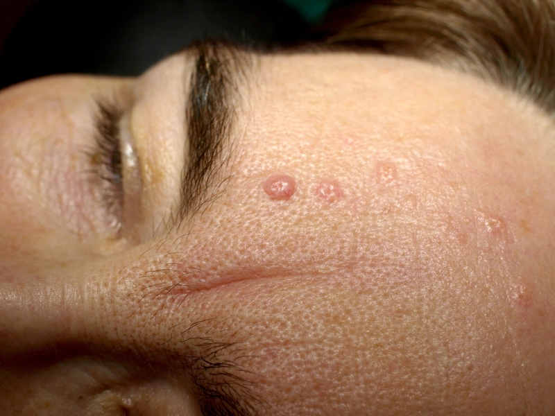
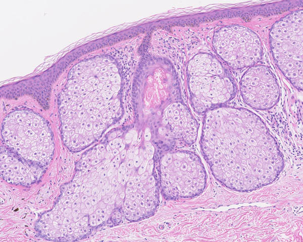 Histology
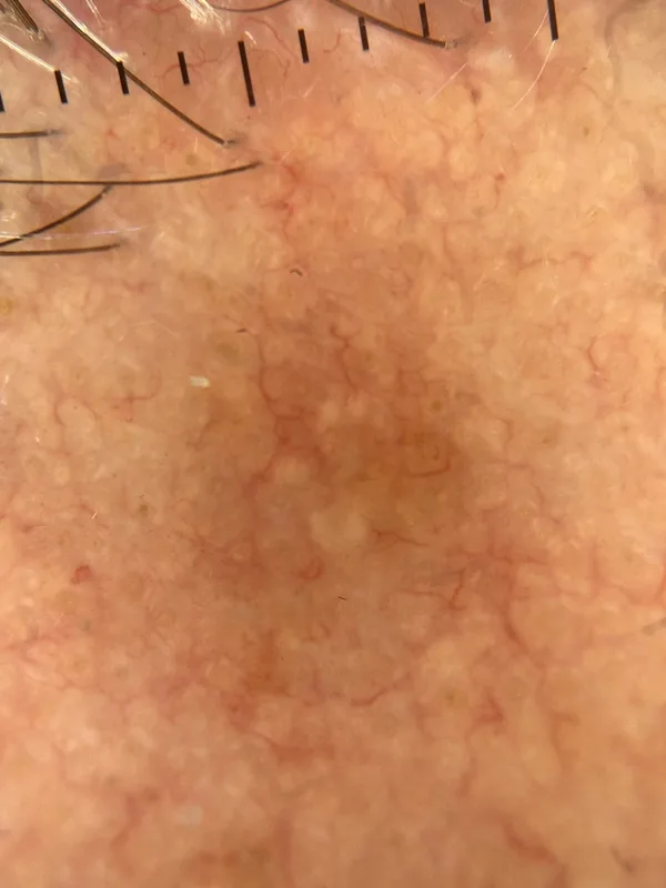 Dermoscopy
Definition
Sebaceous hyperplasia is a benign enlargement of sebaceous glands in which yellowish lobules surround a central hair follicle; it is classed as a benign hair follicle tumour.
Occurrence & epidemiology
It occurs in middle-aged and older people and may be more prevalent under immunosuppression, for example after organ transplantation.
Clinical features
- Single or multiple small yellowish papules up to 3 mm with a central hair follicle, mainly on the forehead and cheeks.
- Often prominent fine vessels, best seen on dermoscopy.
Diagnosis
- Dermoscopy: yellowish lobules around a central follicular opening and prominent vessels.
- The main differential is nodular basal cell carcinoma (arborising vessels, pearly sheen); biopsy if uncertain.
Differential diagnoses
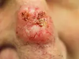
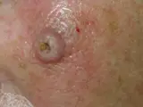
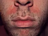
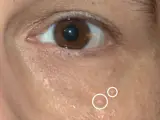
Image credits for the thumbnails
- Trichoepithelioma: Klaus D. Peter, Wiehl, Germany, “Trichoepithelioma”, CC BY 3.0 de, via Wikimedia Commons · CC BY 3.0 de · Source
- Basal cell carcinoma: Bin im Garten, “Basaliom am Nasenrücken 2”, CC BY-SA 3.0, via Wikimedia Commons · CC BY-SA 3.0 · Source
- Keratoacanthoma: Klaus D. Peter, Wiehl, Germany, “Spinaliom”, CC BY 3.0 DE, via Wikimedia Commons · CC BY 3.0 DE · Source
- Seborrhoeic dermatitis: Roymishali, “Seborrhoeic dermatitis highres”, CC BY-SA 3.0, via Wikimedia Commons · CC BY-SA 3.0 · Source
- Syringoma: Mark A. Taff, “Syringoma”, CC BY-SA 3.0 US, via Wikimedia Commons · CC BY-SA 3.0 US · Source
- Milia (milium): Serephine, “Newborn Milia (Milk Spots)”, CC0, via Wikimedia Commons · CC0 · Source
Treatment principles
- Sebaceous hyperplasia is harmless and does not require treatment.
- If desired for cosmetic reasons, lesions can be removed by electrocautery, laser, curettage or cryotherapy, although recurrence and scarring are possible.
- Differentiation from basal cell carcinoma is important, by dermoscopy or histology if in doubt.
Specific treatment regimens with drugs and doses are covered in the DermaFuchs app.
Keep learning in the app
References (selection)
Cross-references
Keep learning
Keep learning with flashcards, exam questions and spot diagnoses in the DermaFuchs app – free, in your browser or as an app.
Related pages: Trichoepithelioma · Basal cell carcinoma · Keratoacanthoma
Note: This page is intended for medical education and does not replace diagnosis or treatment decisions in individual cases.