Infantile hemangioma
Exam relevance: in 1 of more than 50 board exam reports · rank 93
- Synonyms
- Blutschwamm, Säuglingshämangiom, strawberry mark, infantile haemangioma
- Specialty
- Dermatology · Skin tumours
- Images
- Clinical 6 · Histology 1
- In the app
- 4 flashcards
- Last updated
- 10/2026 · Dr. Pascal Bafteh
Contents
Images (7)
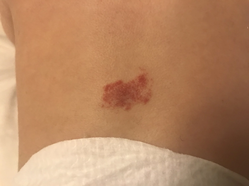
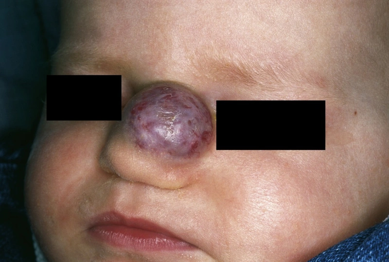
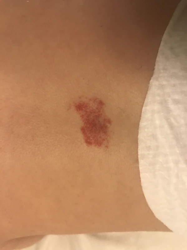
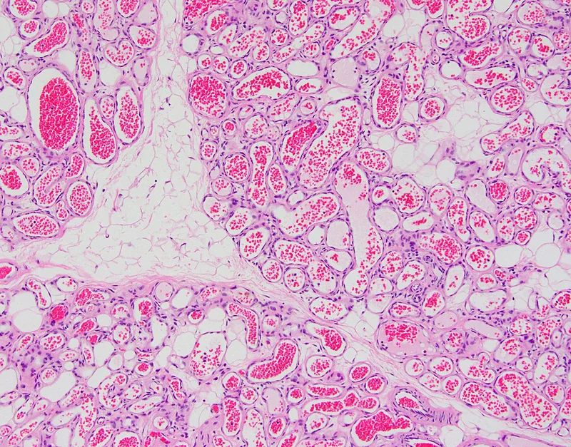 Histology
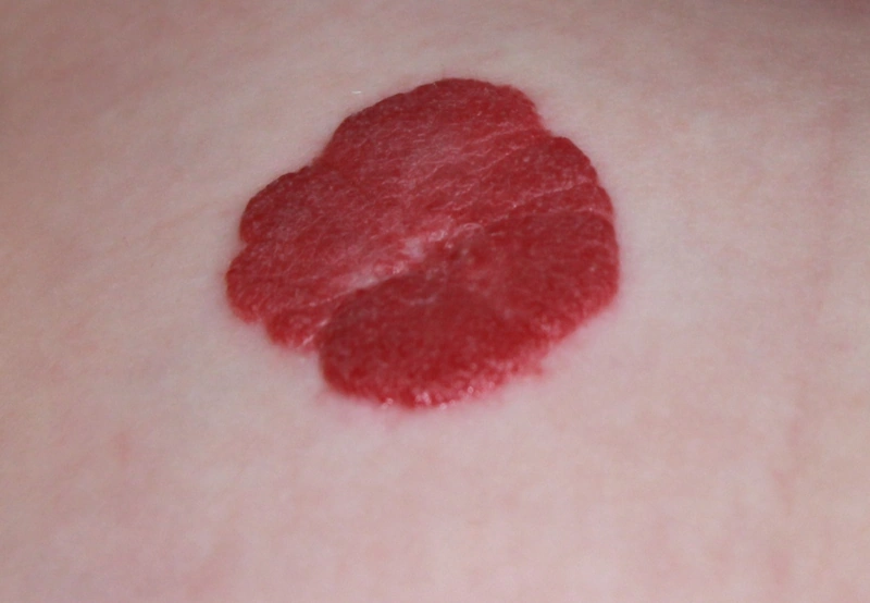
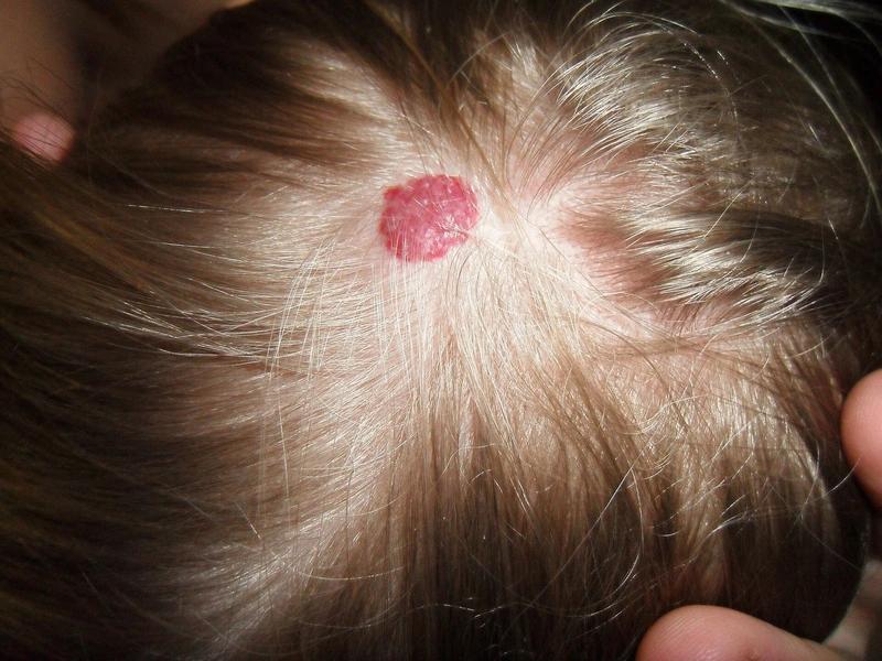
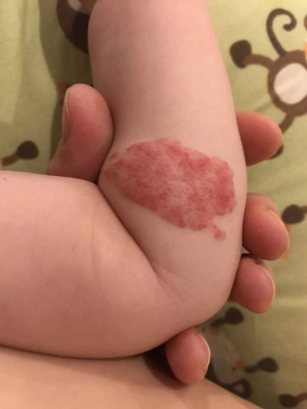
Definition
Overview: ISSVA Classification 2018
- Tumors: Infantile hemangioma (IH), congenital hemangiomas (RICH/PICH/NICH), pyogenic granuloma, angiosarcoma — Characteristic: proliferative, GLUT1±, mitotically active
- Malformations: Nevus flammeus / Port-Wine Stain, AVM, venous / lymphatic / combined — Characteristic: NO proliferation, grows with patient, no spontaneous regression
Infantile Capillary Hemangioma (IH)
Epidemiology & Pathogenesis
- Incidence: 4–5% of all infants, premature infants under 1000 g up to 30%, sex F: M = 3: 1.
- Pathogenesis: The cause is angiogenesis defect (VEGF, placental hypoxia) — IH endothelial cells express GLUT1 + (together with placental endothelia), indicating placental embolization or stem cell differentiation.
Diagnostics
- Clinical + Ultrasound / Doppler (hypervascular lobular architecture), for deep involvement MRI with contrast medium.
- GLUT1 immunohistochemistry: for clinically unclear findings — GLUT1+ = IH; GLUT1− (negative) = congenital hemangioma (RICH/PICH/NICH) or malformation.
Complications
- General: Ulceration (10–15%, pain!), secondary infection, bleeding, scarring residual changes.
Associated Syndromes
- PHACES: Acronym = Post. Fossa + Hemangioma + Arterial + Cardiac + Eye + Sternal — MRI skull + echo + ophthalmological evaluation obligatory.
- PELVIS / SACRAL / LUMBAR: segmental ulcerated lumbosacral hemangiomas + urogenital + spinal malformations → MRI spine + pelvis obligatory.
- Disseminated Hemangiomatosis: ≥ 5 cutaneous hemangiomas → Ultrasound screening liver / GI tract for visceral hemangiomas.
Differential diagnoses
 Pyogenic granuloma
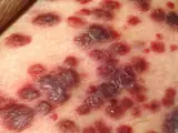 Kaposi's sarcoma
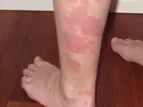 Port-wine stain (naevus flammeus)
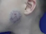 Congenital haemangioma (RICH, PICH, NICH)
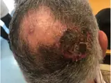 Cutaneous angiosarcoma
- Haemangioma
Image credits for the thumbnails
- Pyogenic granuloma: kilbad, “Pyogenic granuloma on a finger-1”, CC BY 3.0, via Wikimedia Commons · CC BY 3.0 · Source
- Kaposi's sarcoma: unknown author, “Kaposi's Sarcoma”, Public Domain, via Wikimedia Commons · Public Domain · Source
- Port-wine stain (naevus flammeus): Dr. Wonko, CC BY-SA 4.0, via Wikimedia Commons · CC BY-SA 4.0 · Source
- Congenital haemangioma (RICH, PICH, NICH): SafinDinar, “Врожденная не инволюционирующая гемангиома (Noninvoluting congenital hemangioma) (NICH)”, CC BY-SA 4.0, via Wikimedia Commons · CC BY-SA 4.0 · Source
- Cutaneous angiosarcoma: Messias H, Martins M, Zagalo C et al. (Cancer Rep (Hoboken) 2022), CC BY 4.0, via PubMed Central (PMC9575495) · CC BY 4.0 · Source
Treatment principles
- Small haemangiomas at low-risk sites without functional impairment can be managed by watchful waiting, as they regress spontaneously.
- Treatment is indicated mainly for ulceration, threatened function (e.g. periocular, periorificial, airway) or risk of disfigurement; the decision should be made early in the proliferative phase.
- First-line therapy is systemic beta-blockers, or topical beta-blockers for small superficial lesions; pulsed dye laser and cryotherapy are adjuncts, and glucocorticoids are now second-line.
Guideline: AWMF guideline register (no. 006-100)
Specific treatment regimens with drugs and doses are covered in the DermaFuchs app.
Keep learning in the app
References (selection)
Cross-references
Keep learning
Keep learning with flashcards, exam questions and spot diagnoses in the DermaFuchs app – free, in your browser or as an app.
Related pages: Pyogenic granuloma · Kaposi's sarcoma · Port-wine stain (naevus flammeus)
Note: This page is intended for medical education and does not replace diagnosis or treatment decisions in individual cases.