Necrolytic migratory erythema
- Synonyms
- glucagonoma
- Specialty
- Dermatology · Metabolic skin disorders
- Images
- Clinical 4
- In the app
- 2 flashcards
- Last updated
- 10/2026 · Dr. Pascal Bafteh
Contents
Images (4)
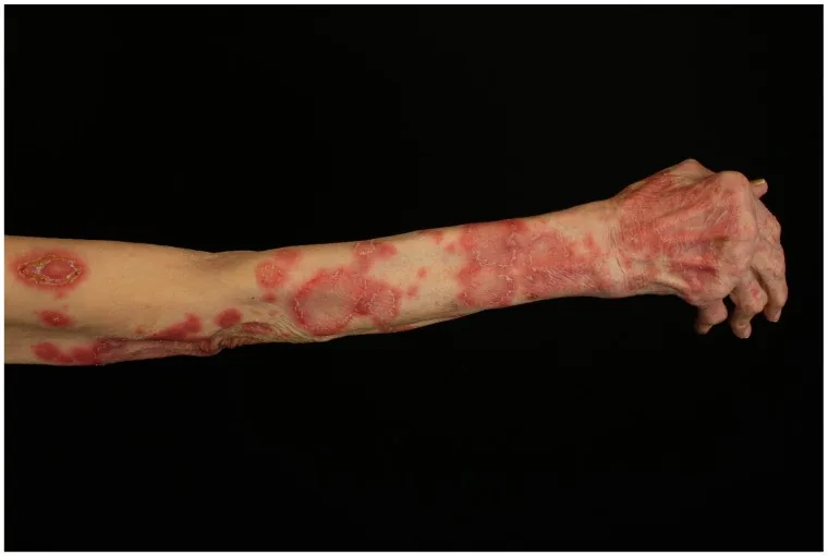
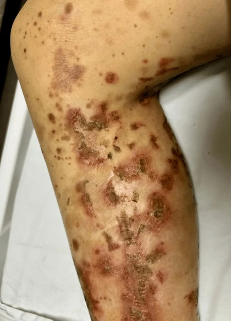
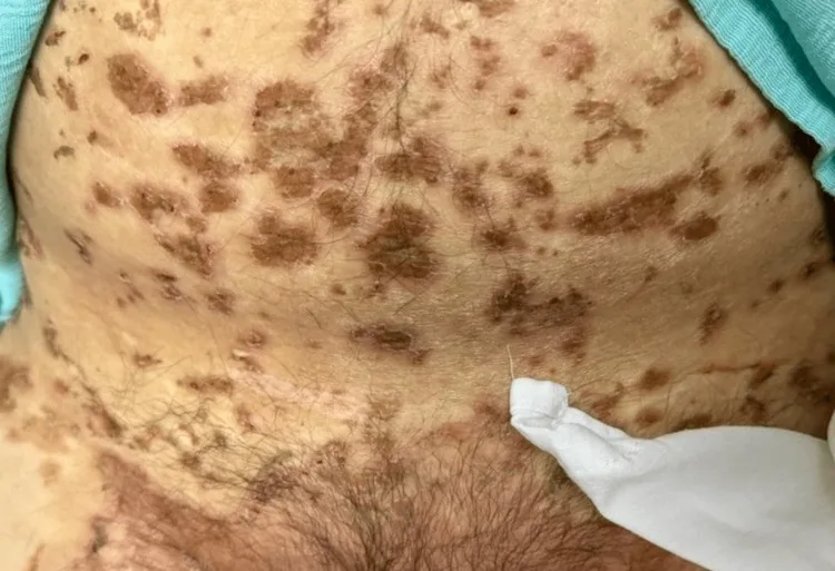
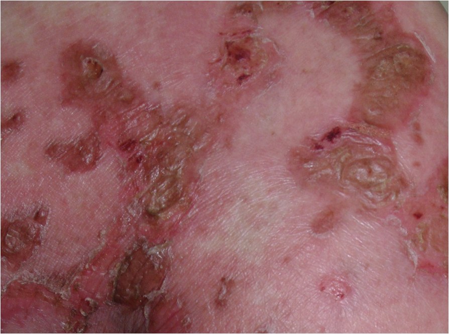
Definition
Definition & Pathogenesis
- Necrolytic migratory erythema (NME) = obligate paraneoplastic dermatosis of the glucagonoma (neuroendocrine pancreatic tumor, α-cell tumor), described as glucagonoma syndrome by Mallinson et al. in 1974.
- Pathogenesis: Mechanism is hyperglucagonemia + hypoaminoacidemia + zinc/niacin/tryptophan deficiency → epidermal necrolysis with defect of keratinocyte energy supply.
- Tumor: Causative tumor is glucagonoma (neuroendocrine α-cell pancreatic tumor; > 50 % already metastatic at diagnosis, usually liver).
- Pseudo-glucagonoma syndrome: NME without glucagonoma in liver cirrhosis, pancreatitis, celiac disease, malabsorption (pseudo-NME) — important DD.
Clinical features
Clinical Presentation
- Skin triad: Characteristic efflorescences are migratory, annular/circinate confluent erythemas + central bullae/erosions + marginal scaling + postinflammatory hyperpigmentation; perigenital + perianal + intertrigines + acra.
- Mucosa: glossitis (red-smooth tongue), angular cheilitis, stomatitis.
- System signs: Classic 4-D tetrad is Dermatitis (NME) + Diabetes mellitus + Deep vein thrombosis + Depression (+ weight loss, diarrhoea).
- Laboratory triad: hyperglucagonemia + hypoaminoacidemia + normochromic normocytic anemia.
- Thrombosis risk: ↑ in glucagonoma (paraneoplastic hypercoagulability).
Diagnosis
Diagnostics
- Skin biopsy: Histological hallmark is necrolytic upper epidermal layer ("pale upper epidermis" with ballooning + confluence of necrotic keratinocytes) + parakeratosis; similar to pellagra / acrodermatitis enteropathica / necrolytic acral erythema.
- Laboratory: Guiding lab findings are plasma glucagon ↑↑ (often many times normal) + amino acid levels ↓ + zinc/niacin often ↓ + hyperglycemia + normochromic anemia.
- Genetics: rule out MEN-1 (multiple endocrine neoplasia type 1) in younger patients / family history.
Differential diagnoses
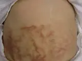 Erythema ab igne
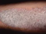 Psoriasis vulgaris (plaque psoriasis)
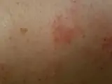 Nummular eczema (discoid eczema)
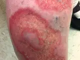 Necrobiosis lipoidica
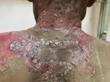 Pemphigus vulgaris
Image credits for the thumbnails
- Erythema ab igne: James Heilman, MD, “Hotbottlerash”, CC BY-SA 3.0, via Wikimedia Commons · CC BY-SA 3.0 · Source
- Psoriasis vulgaris (plaque psoriasis): MediaJet, “An Arm Covered With Plaque Type Psoriasis”, CC BY-SA 3.0, via Wikimedia Commons · CC BY-SA 3.0 · Source
- Nummular eczema (discoid eczema): Ifnord, “Nummular dermatitits”, CC BY-SA 4.0, via Wikimedia Commons · CC BY-SA 4.0 · Source
- Necrobiosis lipoidica: Warfieldian, “Necrobiosislipoidica”, CC BY-SA 3.0, via Wikimedia Commons · CC BY-SA 3.0 · Source
- Pemphigus vulgaris: Mohammad2018, CC BY-SA 4.0, via Wikimedia Commons, cropped · CC BY-SA 4.0 · Source
Treatment principles
- Treating the underlying glucagonoma is crucial; surgical tumour resection is the only curative option but is often not possible because liver metastases are frequently present at diagnosis.
- Somatostatin analogues, which inhibit glucagon secretion, provide symptomatic relief; substitution of amino acids, trace elements and essential fatty acids often improves the skin lesions rapidly.
- Metastatic disease is managed by the interdisciplinary tumour board; because of the increased thrombotic tendency, thromboprophylaxis should be considered.
Specific treatment regimens with drugs and doses are covered in the DermaFuchs app.
Keep learning in the app
References (selection)
Cross-references
Mentioned in
Keep learning: test yourself
One question from the DermaFuchs app on Necrolytic migratory erythema – tap an answer.
Question 1 of 1
What type of pancreatic tumor is Glucagonoma?
Answer
Correct: Pancreatic α-cell tumor. Glucagonoma is a pancreatic α-cell tumor, which can cause necrolytic migratory erythema.
More questions like these – with explanations and spaced repetition – in the app.
Note: This page is intended for medical education and does not replace diagnosis or treatment decisions in individual cases.