Necrobiosis lipoidica
- Specialty
- Dermatology · Metabolic skin disorders
- Images
- Clinical 2 · Histology 1
- In the app
- 5 flashcards
- Last updated
- 10/2026 · Dr. Pascal Bafteh
Contents
Images (3)
 Histology
Definition
Definition & Pathogenesis
- Necrobiosis lipoidica (NL, formerly Necrobiosis lipoidica diabeticorum) = idiopathic / diabetes-associated granulomatous dermatosis with collagen degeneration + lipid deposition.
- Pathogenesis: Suspected mechanisms are microangiopathy + immune complex vasculitis + disturbed collagen metabolism + increased TGF-β / adhesion molecule expression + Th1-driven granuloma formation.
- Diabetes association: Correct regarding the association NL ↔ diabetes is approx. 11–65 % of NL patients have diabetes (type 1 > type 2), but only < 0.3–1 % of diabetics develop NL — diabetes control does NOT correlate with course of skin lesions.
- Associated diseases: thyroid autoimmunity, inflammatory bowel diseases, rheumatoid arthritis, sarcoidosis comorbidity discussed.
Clinical features
Clinical Presentation
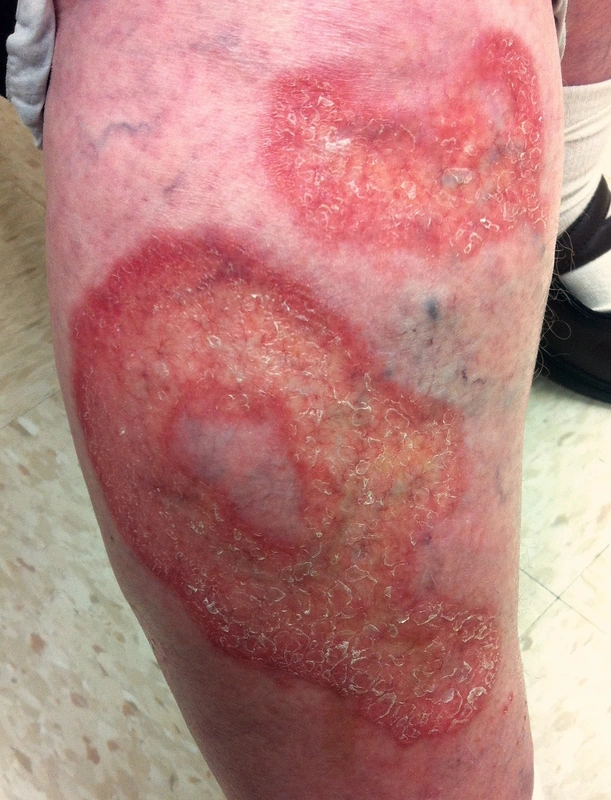
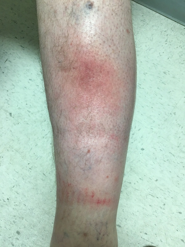
- Location: Predilection is pretibial bilateral (> 85 %), less often thigh, forearm, hands, scalp.
- Morphology: Classic efflorescence is sharply demarcated, yellowish-orange atrophic plaques with livid-erythematous border + visible telangiectasias ("waxy" atrophy).
- Ulceration: Ulceration risk is approx. 25–35 % in course, often painful, poorly healing, banal-trauma-associated.
- Symptoms: long asymptomatic, later pain / hypoesthesia (nerve fiber destruction in plaque), pruritus possible.
- Course: chronic progressive, spontaneous remission rare (< 20 %).
Diagnosis
Diagnostics
- Clinical usually diagnostic (typical yellowish-atrophic pretibial plaques).
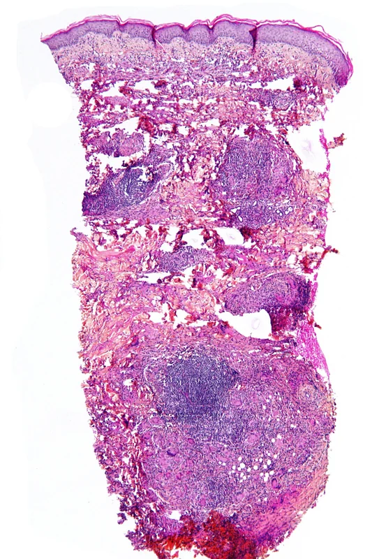
- Histology: Pathognomonic are palisading granulomas around necrobiosis zones (degenerated collagen bundles) throughout the entire dermis, "horizontally layered/tiered", plasma cell involvement, possibly lipid deposits.
- Laboratory: fasting blood glucose / oGTT / HbA1c (diabetes screening), thyroid parameters, possibly ANA / RF / ACE.
- In ulceration: biopsy of border → SCC exclusion (rarely squamous cell carcinoma on chronic NL).
Differential diagnoses
 Granuloma annulare
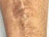 Diabetic dermopathy (shin spots)
 Morphea
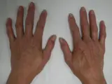 Limited systemic sclerosis (CREST syndrome)
 Cutaneous sarcoidosis
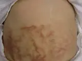 Erythema ab igne
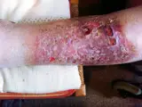 Venous leg ulcer
Image credits for the thumbnails
- Granuloma annulare: Kreuter et al., “Generalized granuloma annulare”, CC BY-SA 2.0, via Wikimedia Commons · CC BY-SA 2.0 · Source
- Diabetic dermopathy (shin spots): Abate, Aroucha, Nóbrega et al. (2025), “Cutaneous manifestations of diabetes mellitus: a narrative review”, CC BY 4.0, via Wikimedia Commons · CC BY 4.0 · Source
- Morphea: Leith C Jones at English Wikipedia, “MercMorphea”, CC BY 3.0, via Wikimedia Commons · CC BY 3.0 · Source
- Limited systemic sclerosis (CREST syndrome): James Heilman, MD, “CREST syndrome (calcinosis and sclerodactyly)”, CC BY-SA 4.0, via Wikimedia Commons · CC BY-SA 4.0 · Source
- Cutaneous sarcoidosis: Nowack et al., “Cutaneous findings in systemic sarcoidosis”, CC BY-SA 2.0, via Wikimedia Commons · CC BY-SA 2.0 · Source
- Erythema ab igne: James Heilman, MD, “Hotbottlerash”, CC BY-SA 3.0, via Wikimedia Commons · CC BY-SA 3.0 · Source
- Venous leg ulcer: Abdullah Sarhan, “Chronisch-venöse Insuffizienz Stadium (III)”, CC BY-SA 4.0, via Wikimedia Commons · CC BY-SA 4.0 · Source
Treatment principles
- General measures include avoiding minor trauma, smoking cessation and good diabetes control, although this does not correlate with the course of the skin disease.
- First-line treatment is potent topical glucocorticoids under occlusion or intralesionally at the active border, and topical calcineurin inhibitors; phototherapy (e.g. PUVA) is an adjunct.
- Refractory disease requires systemic therapies; ulcerated lesions need structured wound management and, if necessary, skin grafting.
Specific treatment regimens with drugs and doses are covered in the DermaFuchs app.
Keep learning in the app
References (selection)
Cross-references
Keep learning
Keep learning with flashcards, exam questions and spot diagnoses in the DermaFuchs app – free, in your browser or as an app.
Related pages: Granuloma annulare · Diabetic dermopathy (shin spots) · Morphea
Note: This page is intended for medical education and does not replace diagnosis or treatment decisions in individual cases.