Skin cysts (epidermoid cyst, milia & more)

Exam relevance: in 3 of more than 50 board exam reports · rank 45

Synonyms
Atherom, Epidermalzyste, Milien, skin cyst, epidermoid cyst, sebaceous cyst, atheroma, milia
Specialty
Dermatology · Skin appendages
Images
Clinical 7 · Histology 4
In the app
2 flashcards
Last updated
10/2026 · Dr. Pascal Bafteh
Contents
  1. Images (11)
  2. Classification
  3. Treatment principles
  4. Keep learning in the app
  5. References (selection)
  6. Cross-references

Images (11)

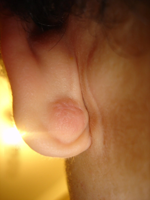
Skin cysts (epidermoid cyst, milia & more) – clinical photo: firm, skin-coloured nodule on the earlobe
firm, skin-coloured nodule on the earlobeunknown (Commons user), “Epidermal Cyst ear”, CC BY-SA 3.0, via Wikimedia Commons · CC BY-SA 3.0 · Source
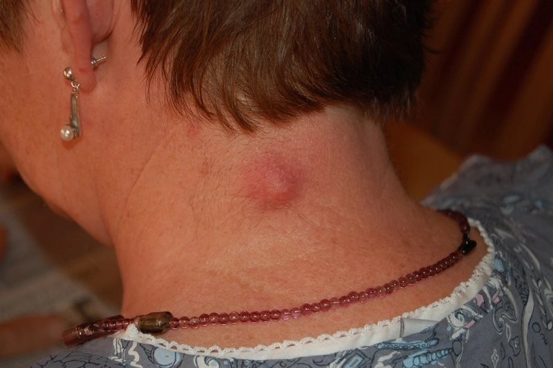
Skin cysts (epidermoid cyst, milia & more) – clinical photo: erythematous, tender nodule on the neck with a central punctum
erythematous, tender nodule on the neck with a central punctumSteven Fruitsmaak, “Inflamed epidermal inclusion cyst”, CC BY-SA 3.0, via Wikimedia Commons · CC BY-SA 3.0 · Source
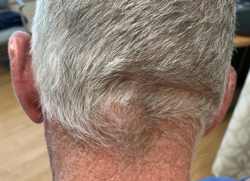
Skin cysts (epidermoid cyst, milia & more) – clinical photo: flat, skin-coloured subcutaneous swelling on the lower back of the head
flat, skin-coloured subcutaneous swelling on the lower back of the headDermanonymous, “Epidermal Inclusion Cyst”, CC BY-SA 4.0, via Wikimedia Commons · CC BY-SA 4.0 · Source
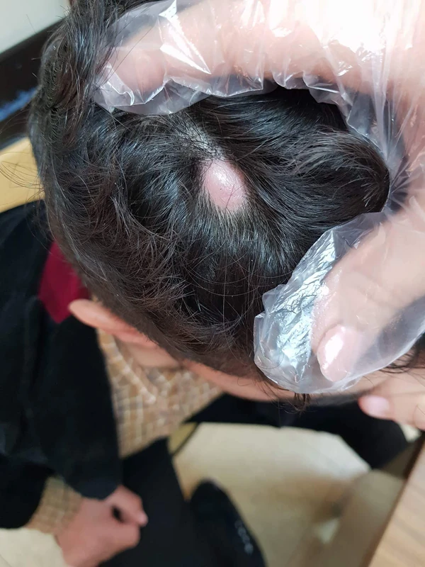
Skin cysts (epidermoid cyst, milia & more) – clinical photo: scalp: solitary, smooth, dome-shaped, skin-colored to pink nodule on the vertex with a hairless surface
scalp: solitary, smooth, dome-shaped, skin-colored to pink nodule on the vertex with a hairless surfaceMasryyy, “Sbecous cyst new photo for diagnosis”, CC BY-SA 4.0, via Wikimedia Commons · CC BY-SA 4.0 · Source
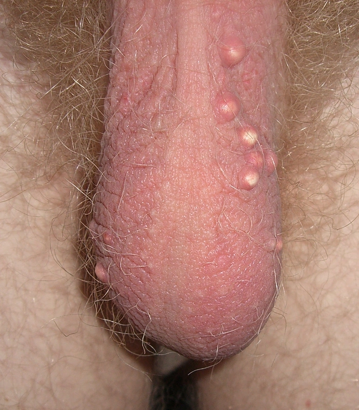
Skin cysts (epidermoid cyst, milia & more) – genital area – clinical photo: several firm yellowish-white papules and nodules
several firm yellowish-white papules and nodules (genital area)Empressario, “Scrotal epidermoid cysts”, Public Domain, via Wikimedia Commons · Public domain · Source
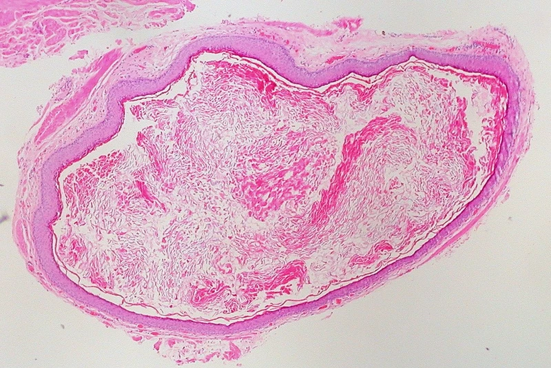
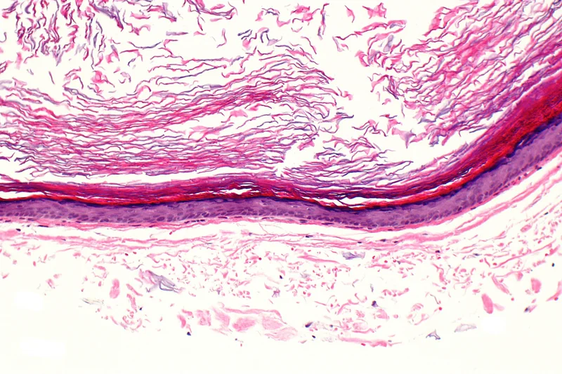
Skin cysts (epidermoid cyst, milia & more) – histology (H&E): overview of a unilocular dermal cyst with thin epithelial wall and luminal keratinHistology
histology (H&E): overview of a unilocular dermal cyst with thin epithelial wall and luminal keratinEd Uthman from Houston, TX, USA, “Epidermal Inclusion Cyst (5517587941)”, CC BY 2.0, via Wikimedia Commons · CC BY 2.0 · Source
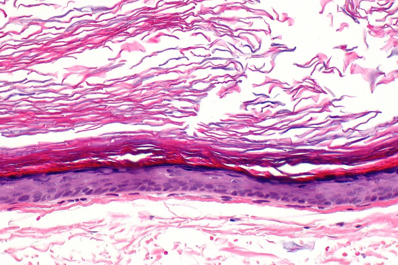
Skin cysts (epidermoid cyst, milia & more) – histology (H&E): squamous epithelial cyst wall with granular layer, laminated basket-weave keratin in the lumenHistology
histology (H&E): squamous epithelial cyst wall with granular layer, laminated basket-weave keratin in the lumenNephron, “Epidermal inclusion cyst -- high mag”, CC BY-SA 3.0, via Wikimedia Commons · CC BY-SA 3.0 · Source
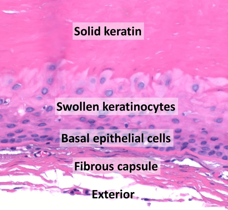
Skin cysts (epidermoid cyst, milia & more) – histology (H&E, labelled): compact, homogeneously eosinophilic keratin without a granular layer, abrupt trichilemmal keratinisation, swollen pale keratinocytes in the uppermost …Histology
histology (H&E, labelled): compact, homogeneously eosinophilic keratin without a granular layer, abrupt trichilemmal keratinisation, swollen pale keratinocytes in the uppermost …Mikael Häggström, M.D., “Histopathology of trichilemmal cyst - annotated”, CC0, via Wikimedia Commons · CC0 · Source
Skin cysts (epidermoid cyst, milia & more) – histology (H&E): a cyst wall of stratified keratinising squamous epithelium with a compact horn lamella facing the lumen and lamellated keratin within it; the granular layer is only …Histology
histology (H&E): a cyst wall of stratified keratinising squamous epithelium with a compact horn lamella facing the lumen and lamellated keratin within it; the granular layer is only …Nephron, “Epidermal inclusion cyst -- intermed mag”, CC BY-SA 3.0, via Wikimedia Commons · CC BY-SA 3.0 · Source
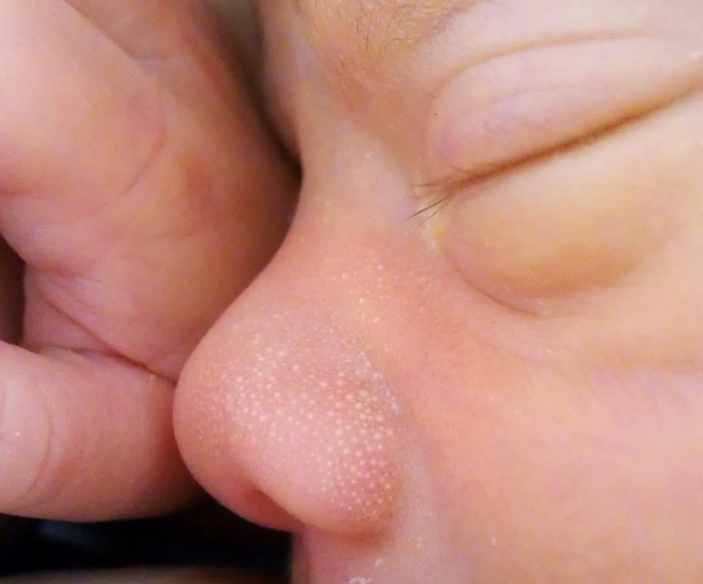
Skin cysts (epidermoid cyst, milia & more) – face, baby – clinical photo: white, pinhead-sized cysts on the nose of a one-week-old infant
white, pinhead-sized cysts on the nose of a one-week-old infant (face)Serephine, CC0 · CC0 · Source
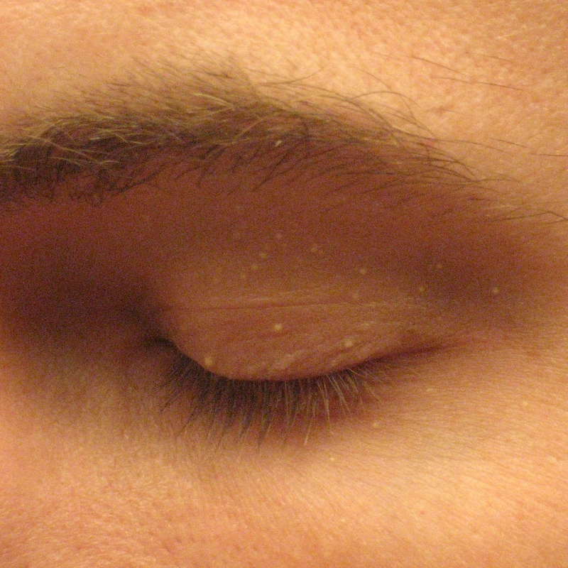
Skin cysts (epidermoid cyst, milia & more) – clinical photo: whitish-yellow, firm-looking retention cysts on the eyelid
whitish-yellow, firm-looking retention cysts on the eyelidOriginal uploader was Silver442n at en.wikipedia, Public domain · Public domain · Source
1 / 11

Classification

Epidermal Cyst (Infundibular Cyst)

  • Most common cutaneous cyst type, lined with keratinizing squamous epithelium.
  • Content: Keratin/horn lamellae. Clinical presentation: subcutaneous, mobile nodule with pore.
  • Complication: Rupture → foreign body reaction.
  • Ultrasound (high-frequency linear probe, approx. 7–15 MHz): sharply demarcated, hypoechoic lesion with posterior acoustic enhancement and lateral edge shadowing — the classic proof of a cyst; the lesion itself is avascular on colour Doppler.

Dermoid Cyst

  • Wall of epidermis with skin appendages (hair, sebaceous and sweat glands).
  • Location: Lateral eyebrow/midline. Congenital, usually discovered in childhood.

Milia (Milium)

  • Definition: 1–2 mm whitish-yellow, firm epidermal keratin cysts — essentially a miniature epidermal cyst.
  • Primary milia: no preceding damage, predilection periorbital and cheeks. As milia neonatorum in about 40–50 % of newborns and self-limiting.
  • Distinction: syringoma (skin-coloured to yellowish dermal papules, lower lid), xanthelasma (yellow plaques), sebaceous hyperplasia (central dell) — milia are firm and white, without a central dell.

Associated Syndromes

  • Gardner Syndrome: Epidermoid cysts + desmoid tumors + osteomas + FAP. Cause: Mutation in the APC gene.
  • Gorlin-Goltz Syndrome: Jaw keratocysts + multiple BCCs. Mutation: PTCH1.

Treatment principles

  • Epidermoid cysts are excised completely, including the cyst wall, if symptomatic or at the patient's request, as residual wall fragments lead to recurrence.
  • Inflamed or abscessed cysts are first relieved by incision and drainage and removed definitively only once the inflammation has subsided.
  • Milia can be expressed after a fine incision of the overlying skin.

Specific treatment regimens with drugs and doses are covered in the DermaFuchs app.

Keep learning in the app

In the DermaFuchs app you can learn Skin cysts (epidermoid cyst, milia & more) with flashcards, exam questions and spot-the-diagnosis – free, in your browser or as an app.

Test yourself: Spot-diagnosis quiz with real cases →

In the app: 2 flashcards

Open in browser  Download on the App Store

References (selection)

  1. Gallardo Avila PP, Mendez MD. Milia. In: StatPearls. Treasure Island (FL): StatPearls Publishing; 2023.

Cross-references

Keep learning

Keep learning with flashcards, exam questions and spot diagnoses in the DermaFuchs app – free, in your browser or as an app.

Related pages: Acne fulminans · Hidradenitis suppurativa (acne inversa) · Acne vulgaris

Note: This page is intended for medical education and does not replace diagnosis or treatment decisions in individual cases.