Paget disease of the nipple (mammary Paget disease)
- Synonyms
- Mammärer Morbus Paget, Morbus Paget der Brustwarze, Paget-Karzinom der Brust, Mammary Paget disease, Paget disease of the breast, Paget's disease of the nipple
- Specialty
- Dermatology · Skin tumours
- Images
- Clinical 1 · Histology 1
- In the app
- 1 flashcards
- Last updated
- 10/2026 · Dr. Pascal Bafteh
Contents
Images (2)
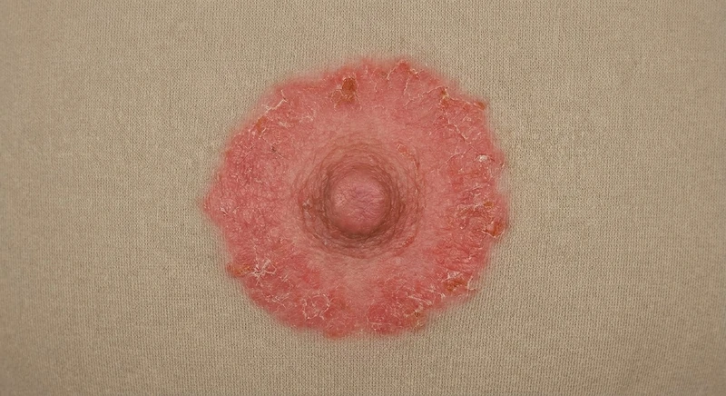 AI illustration
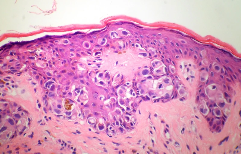 Histology
Definition
Mammary Paget disease is an uncommon form of breast cancer in which tumour cells (Paget cells) infiltrate the epidermis of the nipple and areola. It is nearly always associated with an underlying intraductal carcinoma, in situ or invasive. It is distinct from extramammary Paget disease of the apocrine gland-bearing regions (anogenital skin, axillae).
Occurrence & epidemiology
It accounts for 1–3% of all breast cancers. It mainly affects women in their 50s and 60s, with a wide age range; men are rarely affected.
Aetiopathogenesis
According to the epidermotropic theory, Paget cells arise in an intraductal carcinoma and migrate along the lactiferous ducts into the nipple epidermis. Another theory regards them as malignantly transformed keratinocytes of the nipple skin.
Clinical features
- Usually a unilateral eczema- or psoriasis-like change of the nipple that slowly extends over the areola; a well-defined but irregularly shaped red patch, scaly or crusted, up to 15 cm across.
- Initially asymptomatic, later itchy, burning or painful; the nipple may become thickened or retracted; erosion, bleeding and discharge can occur.
- About 50–60% of patients have a palpable breast lump at presentation.
- A rare pigmented variant is clinically difficult to distinguish from melanoma.
Diagnosis
- Suspected with a longstanding, slowly progressive, unilateral change of the nipple.
- Skin biopsy: typical foamy Paget cells in the epidermis; in the pigmented variant, immunohistochemistry (e.g. CK7, S100, Melan-A, HMB-45) to distinguish them from melanoma cells. A wedge biopsy of the nipple also samples deeper breast tissue.
- Breast imaging (ultrasound, mammography, MRI) to look for the underlying carcinoma and determine its extent.
Differential diagnoses
Keep learning in the app
References (selection)
Cross-references
Note: This page is intended for medical education and does not replace diagnosis or treatment decisions in individual cases.