Zoon balanitis (plasma cell balanitis)
- Synonyms
- Balanitis circumscripta plasmacellularis, Zoon-Balanitis, plasmazelluläre Balanitis, Plasma cell balanitis
- Specialty
- Dermatology · Regional & special dermatoses
- Images
- Clinical 1 · Histology 1
- In the app
- 1 flashcards
- Last updated
- 10/2026 · Dr. Pascal Bafteh
Contents
Images (2)

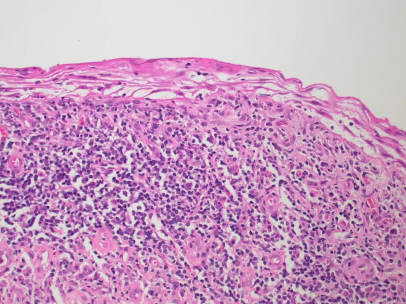 Histology
Definition
Zoon balanitis is a benign, chronic, idiopathic inflammation of the genital mucosa with a plasma cell-rich infiltrate, typically presenting as a well-defined, shiny, red patch on the glans penis. Zoon described it in 1952. Corresponding changes in women are called plasma cell (Zoon) vulvitis.
Occurrence & epidemiology
It predominantly affects uncircumcised middle-aged to older men. Reliable data on incidence and prevalence are lacking; the condition is probably underdiagnosed.
Aetiopathogenesis
The cause is unknown. Chronic mucosal irritation is assumed, related to a “dysfunctional” prepuce with retention of urine and smegma, moisture, friction and heat. The plasma cell infiltrate is polyclonal.
Clinical features
- One or more well-defined, orange-red, shiny, moist macules or slightly raised plaques on the glans, sometimes also on the prepuce.
- “Cayenne pepper spots”: pinpoint brighter red speckles due to microhaemorrhage and haemosiderin deposition.
- Mirror-image lesions sometimes appear on the opposing prepuce (“kissing lesions”); after erosion a rust-coloured stain may remain.
- Usually asymptomatic; occasionally itch, burning, pain or dysuria.
Histology
Early acanthosis and parakeratosis, later epidermal atrophy, erosions and lozenge-shaped keratinocytes. In the upper dermis a band-like infiltrate usually comprising more than 50% plasma cells, with siderophages, extravasated red cells and dilated vessels running vertically to obliquely.
Diagnosis
- Suspected clinically from the typical appearance on the uncircumcised penis.
- Dermoscopy: focal or diffuse orange-yellowish structureless areas (haemosiderin) and curved vessels.
- Biopsy for confirmation, above all to distinguish it from erythroplasia of Queyrat (carcinoma in situ) and other forms of balanitis, such as those due to Candida, lichen planus or psoriasis.
Differential diagnoses
Keep learning in the app
References (selection)
Cross-references
Note: This page is intended for medical education and does not replace diagnosis or treatment decisions in individual cases.