Tinea (dermatophytosis)

Exam relevance: in 12 of more than 50 board exam reports · rank 6

Synonyms
Dermatophytose, Fadenpilzinfektion, ringworm, dermatophytosis, fungal skin infection, tinea
Specialty
Dermatology · Fungal infections
Images
Clinical 16
In the app
11 flashcards
Last updated
10/2026 · Dr. Pascal Bafteh
Contents
  1. Images (16)
  2. Definition
  3. Clinical features
  4. Diagnosis
  5. Treatment principles
  6. Keep learning in the app
  7. References (selection)
  8. Cross-references

Images (16)

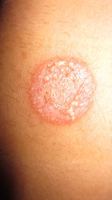
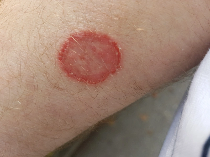
Tinea (dermatophytosis, ringworm) – clinical photo: sharply demarcated, round red plaque with a raised border and yellowish papulovesicles or pustules, mostly at the margin
sharply demarcated, round red plaque with a raised border and yellowish papulovesicles or pustules, mostly at the marginCorina G., “Tinea corporis”, Public Domain, via Wikimedia Commons · Public domain · Source
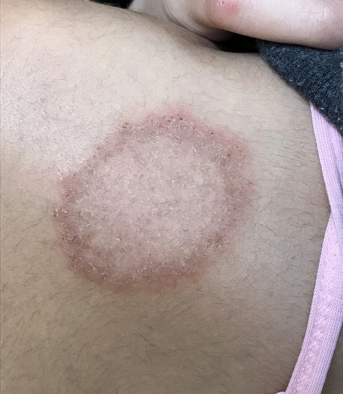
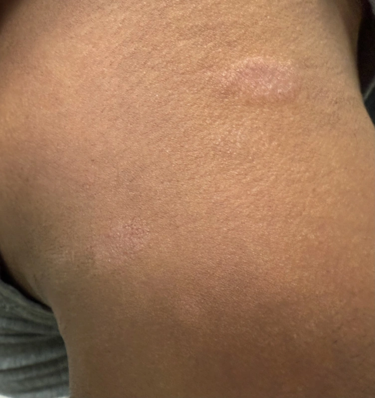
Tinea (dermatophytosis, ringworm) – clinical photo: scaly, annular plaque with active border on the shoulder of a teenager
scaly, annular plaque with active border on the shoulder of a teenagerGzzz, “Dermatophytosis on a teenager's shoulder”, CC BY-SA 4.0, via Wikimedia Commons · CC BY-SA 4.0 · Source
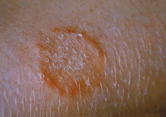
Tinea (dermatophytosis, ringworm) – clinical photo: ring-shaped lesion with a reddish-brown border and scaly centre on the arm
ring-shaped lesion with a reddish-brown border and scaly centre on the armCDC/ Dr. Lucille K. Georg, “Ringworm on the arm, or tinea corporis due to Trichophyton mentagrophytes PHIL 2938 lores”, Public Domain, via Wikimedia Commons · Public domain · Source · CDC PHIL #2938
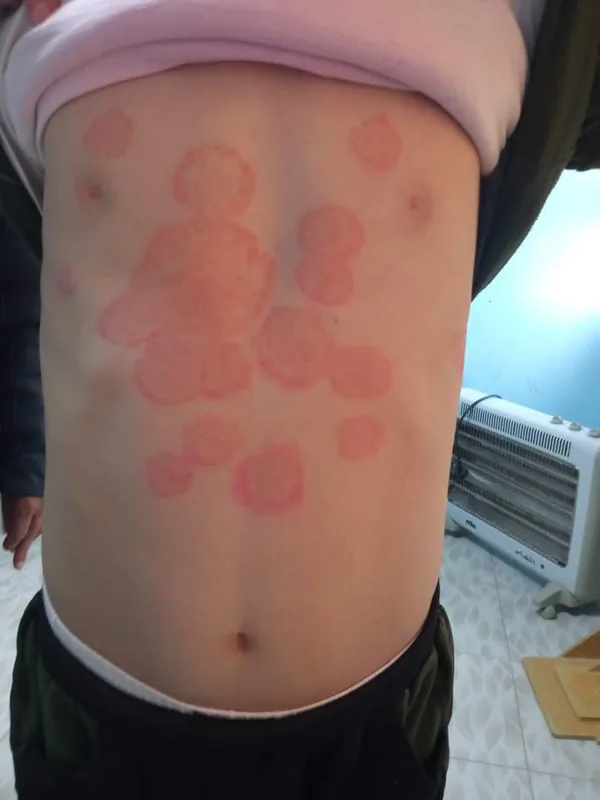
Tinea (dermatophytosis, ringworm) – clinical photo: numerous round, partly confluent red lesions with accentuated borders on the abdomen and chest
numerous round, partly confluent red lesions with accentuated borders on the abdomen and chestMohammad3021, “Tinea corporis”, CC0, via Wikimedia Commons · CC0 · Source
Tinea (dermatophytosis, ringworm) – clinical photo: round lesion with a raised scaly border and clearing centre on the forearm
round lesion with a raised scaly border and clearing centre on the forearmAsurnipal, “Dermatophytosis 20190815-02ASD”, CC BY-SA 4.0, via Wikimedia Commons · CC BY-SA 4.0 · Source
Tinea (dermatophytosis, ringworm) – clinical photo: annular, slightly scaly lesions with a raised border on the nape of the neck
annular, slightly scaly lesions with a raised border on the nape of the neckడా. గన్నవరపు నరసింహమూర్తి, “Tinea corporis over nape of neck”, CC0, via Wikimedia Commons · CC0 · Source
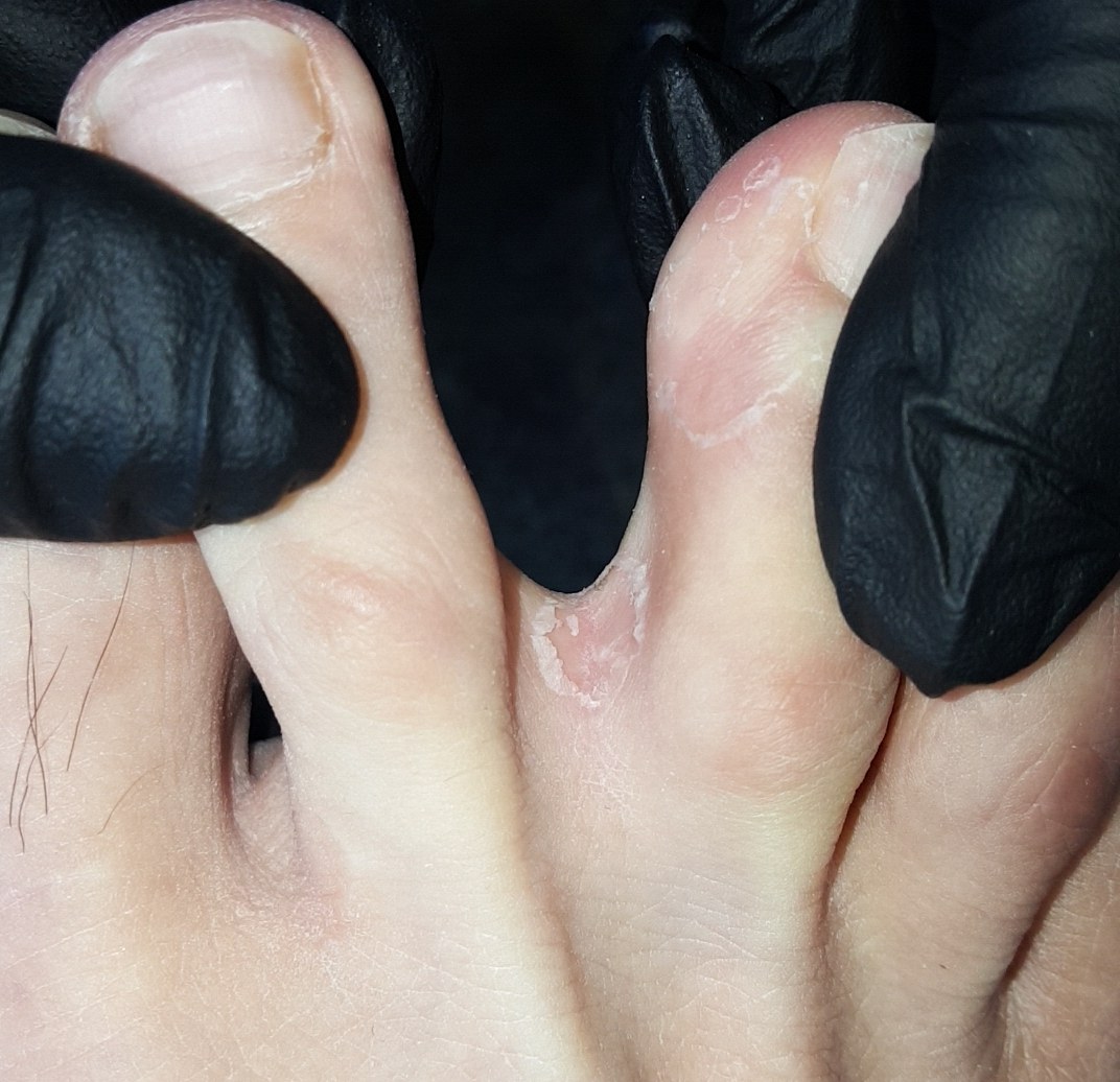
Tinea (dermatophytosis, ringworm) – finger, toe – clinical photo: macerated, scaly skin in the 3rd/4th interdigital space
macerated, scaly skin in the 3rd/4th interdigital space (finger, toe)Tang21237, “Interdigital tinea between the 3rd and 4th interdigital spaces”, CC BY-SA 4.0, via Wikimedia Commons · CC BY-SA 4.0 · Source
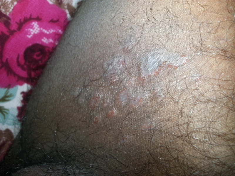
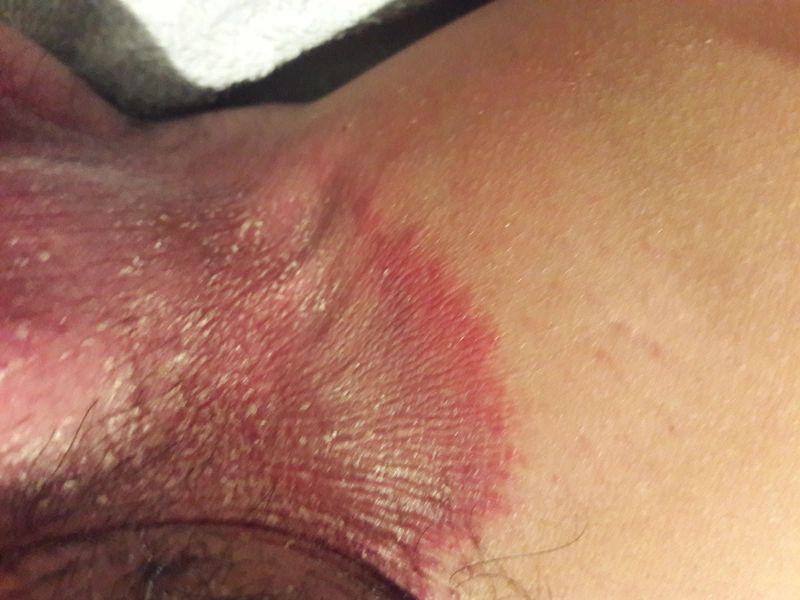
Tinea (dermatophytosis, ringworm) – leg – clinical photo: erythematous, scaly plaque with active, partly pustular border on the groin and inner thigh
erythematous, scaly plaque with active, partly pustular border on the groin and inner thighAjay Kumar Chaurasiya, “Ringworm or Tinea cruris of fungal infection”, CC BY-SA 4.0, via Wikimedia Commons · CC BY-SA 4.0 · Source
Tinea (dermatophytosis, ringworm) – leg – clinical photo: sharply demarcated erythema with a scaly, raised border in the groin, extending onto the inner thigh
sharply demarcated erythema with a scaly, raised border in the groin, extending onto the inner thighRobertgascoign, “Jock Itch”, CC BY-SA 3.0, via Wikimedia Commons · CC BY-SA 3.0 · Source
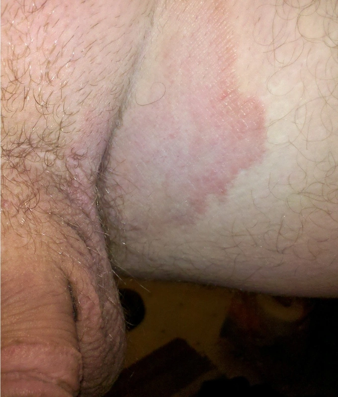
Tinea (dermatophytosis, ringworm) – clinical photo: sharply demarcated, garland-shaped erythematous plaque in the groin
sharply demarcated, garland-shaped erythematous plaque in the groinGerardolagunes, “Jock itch”, CC BY-SA 3.0, via Wikimedia Commons · CC BY-SA 3.0 · Source
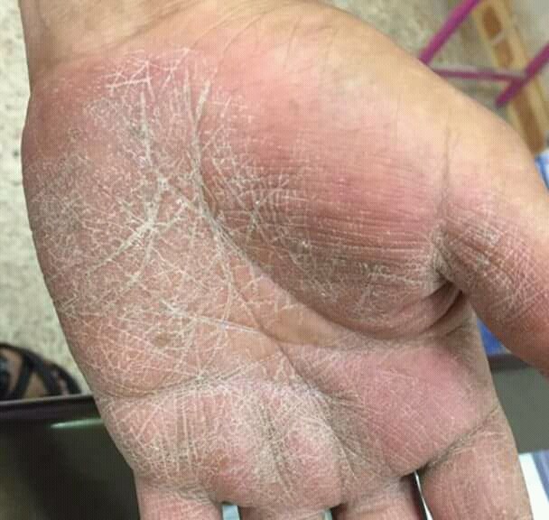
Tinea (dermatophytosis, ringworm) – clinical photo
Tinea (dermatophytosis, ringworm) – clinical photoMohammad2018, “Tinea manum”, CC BY-SA 4.0, via Wikimedia Commons · CC BY-SA 4.0 · Source
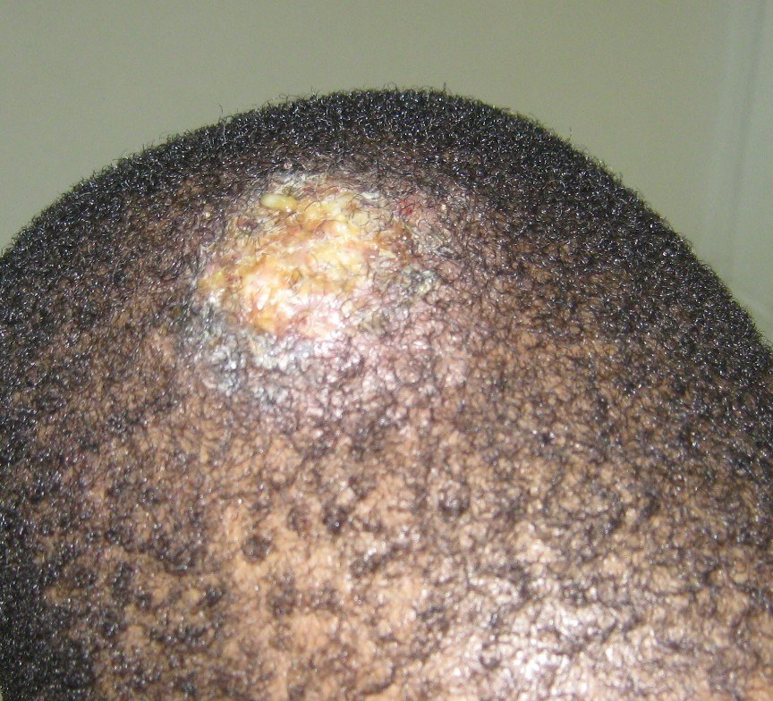
Tinea (dermatophytosis, ringworm) – scalp – clinical photo: painful, boggy, pustular-crusted plaque on the vertex
painful, boggy, pustular-crusted plaque on the vertex (scalp)Grook da oger, “Kérion 01”, CC BY-SA 4.0, via Wikimedia Commons · CC BY-SA 4.0 · Source
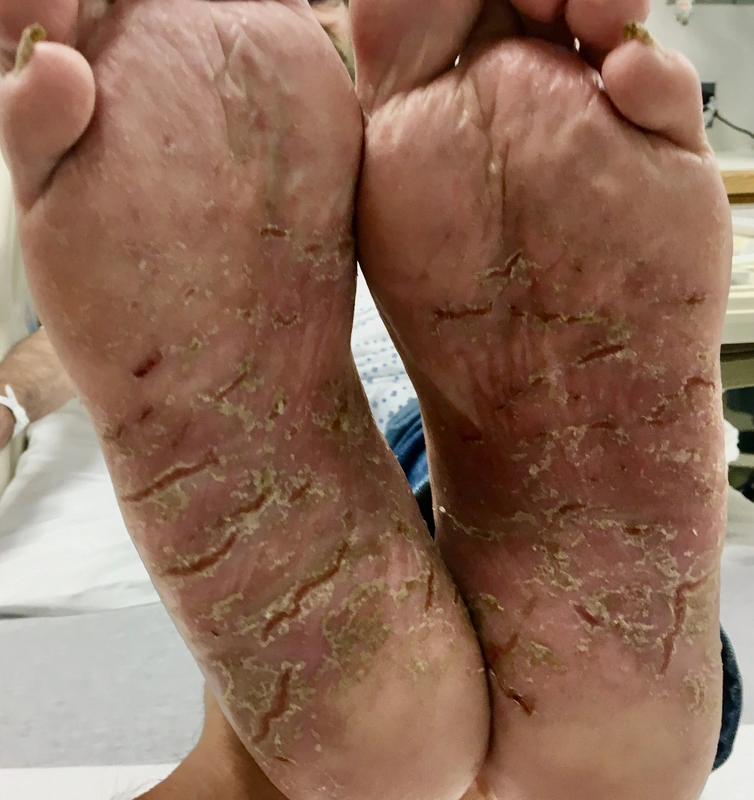
Tinea (dermatophytosis, ringworm) – foot – clinical photo: diffuse hyperkeratosis and fine scaling of both soles
diffuse hyperkeratosis and fine scaling of both soles (foot)డా. గన్నవరపు నరసింహమూర్తి, “Plantar athlete's foot (moccasin foot), tinea pedis”, CC0, via Wikimedia Commons · CC0 · Source
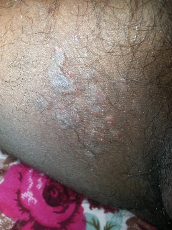
Tinea (dermatophytosis, ringworm) – leg – clinical photo: annular plaque with a scaly active border and central clearing on the thigh in darker skin
annular plaque with a scaly active border and central clearing on the thigh in darker skinAjay Kumar Chaurasiya, “Ringworm or Tinea cruris of fungal infection”, CC BY-SA 4.0, via Wikimedia Commons · CC BY-SA 4.0 · Source
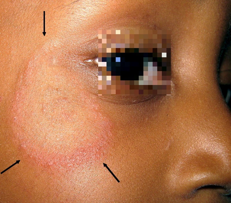
Tinea (dermatophytosis, ringworm) – clinical photo: Tinea faciei: annular lesion on the cheek below the eye with marginal scaling (arrows)
Tinea faciei: annular lesion on the cheek below the eye with marginal scaling (arrows)Grook Da Oger, “Mycose peau glabre - Dermatophytosis”, CC BY-SA 3.0 · CC BY-SA 3.0 · Source
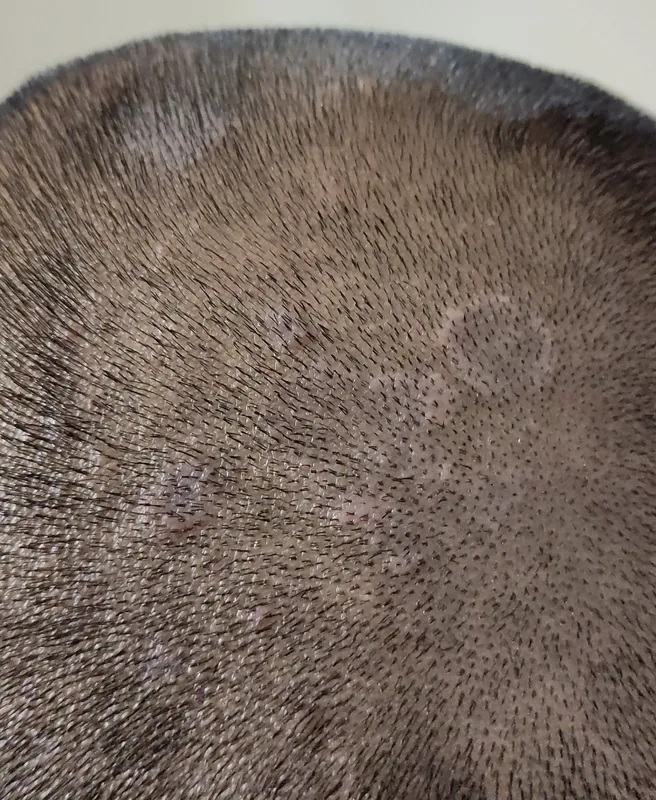
Tinea (dermatophytosis, ringworm) – clinical photo: Tinea capitis: round, finely scaling patches with thinned hair on the closely shaved scalp
Tinea capitis: round, finely scaling patches with thinned hair on the closely shaved scalpVis M, “Fungal infection on head scalp”, CC BY-SA 4.0, via Wikimedia Commons · CC BY-SA 4.0 · Source
1 / 16

Definition

Tinea corporis

  • Clinical features: annular sharply demarcated erythematosquamous plaques with active border and central clearing, often pruritus
  • Pathogens: mainly T. rubrum, also T. interdigitale/mentagrophytes, T. tonsurans, M. canis
  • Affects: Dermatophytes affect skin, hair and nails — including intertriginous regions (tinea inguinalis/cruris).
  • Zoophilic vs. anthropophilic: zoophilic species more aggressive and inflammatory, anthropophilic milder course. Animals often asymptomatic carriers.

Clinical features

Tinea faciei

  • Clinical features: annular erythematosquamous plaques with active border on the face, frequently aggravated by sun exposure — often DD vs. lupus / rosacea / seborrheic eczema.
  • Pathogens: commonly T. rubrum (from feet/nails), M. canis (cat/dog), T. verrucosum (cattle).

Tinea barbae

  • Clinical features: severely inflammatory folliculitis in the beard area with pustules, crusts, beard alopecia — despite marked inflammation often surprisingly little pain or itch; regional LN swelling possible.
  • Pathogens: T. tonsurans, T. mentagrophytes, T. verrucosum.
  • Risk factors: animal contact, shaving trauma, contact sports.

Tinea cruris / pubogenitalis

  • Clinical features: sharply demarcated erythematosquamous plaques in the inguinal/pubic area with active border and severe pruritus; DD candida intertrigo: satellite papules + macerated coatings without active rim.
  • Pathogens: most commonly T. rubrum and Epidermophyton floccosum, less often T. mentagrophytes.

Tinea pedis

  • Clinical forms: Interdigitalis (macerated scaling mainly 4th/5th web space) + Plantaris squamosa (chronic hyperkeratosis in „moccasin distribution") + vesiculobullous form — interdigital form most common.
  • Moccasin type: chronic hyperkeratotic tinea plantaris with fine flour-like scaling on plantae — DD dry foot / psoriasis palmoplantaris.
  • Pathogens: T. rubrum (anthropophilic, most common) and T. interdigitale.

Diagnosis

Specimen Collection

  • Skin: Cleaning with 70% ethanol/isopropanol, scale collection from the active border
  • Nail: Clippings from the transition of healthy/diseased tissue with subungual hyperkeratosis
  • Hair: Epilation of ≥10 hairs or brush sampling

Culture

  • Significance: Gold standard for pathogen specification
  • Setup: Sabouraud and Kimmig agar, possibly with cycloheximide
  • Growth: up to 6 weeks; exception Trichophyton mentagrophytes grows faster, often after 5–7 days
  • By contrast Trichophyton rubrum: slowly (about 2–3 weeks), fluffy white and cotton-like, with a deep red to wine-red reverse.
  • Most important macroscopic criterion besides growth rate and micromorphology: the reverse colour (underside of the colony) — which is why the plate is always inspected from below as well.
  • Further clues: T. mentagrophytes grows faster (about 1 week), granular-powdery, reverse yellow to red-brown · Microsporum canis white and woolly with a yellow-orange reverse · Epidermophyton floccosum khaki yellow-green and velvety, with club-shaped macroconidia and no microconidia.

PCR

  • Characteristics: very sensitive and specific, fast, but without resistance testing
  • Indication: useful for tinea capitis, kerion celsi, and recurrent courses

Wood's Lamp

  • Microsporum canis (tinea capitis): yellow-green fluorescent — classic microsporia detection in children.
  • Trichophyton schoenleinii (favus): blue-white fluorescent.
  • Trichophyton tonsurans / T. verrucosum: no fluorescence — important pitfall, since these common tinea-capitis pathogens stay negative today.
  • Pityriasis versicolor (Malassezia furfur): yellow-orange/greenish fluorescent.

Treatment principles

  • Before treatment, material is taken for direct microscopy and culture, as prior treatment compromises identification of the organism.
  • Localised skin infections are treated topically with antifungals (azoles, allylamines, hydroxypyridones), while involvement of hair, nails or extensive disease is treated systemically, with the agent chosen according to the organism.
  • With zoophilic organisms, the source animal and the environment must be treated; topical glucocorticoids alone mask the picture (tinea incognito).

Guideline: AWMF guideline register (no. 013-002)

Specific treatment regimens with drugs and doses are covered in the DermaFuchs app.

Keep learning in the app

In the DermaFuchs app you can learn Tinea (dermatophytosis) with flashcards, exam questions and spot-the-diagnosis – free, in your browser or as an app.

Test yourself: Spot-diagnosis quiz with real cases →

In the app: 11 flashcards

Open in browser  Download on the App Store

References (selection)

  1. AWMF S1 Tinea 2020
  2. Onychomycosis: An Updated Review
  3. [Tinea capitis in children]
  4. Plewig 2018 Braun-Falco p.265

Cross-references

Keep learning: test yourself

One question from the DermaFuchs app on Tinea (dermatophytosis) – tap an answer.

Question 1 of 1

What is the classic localization and trigger for Majocchi's granuloma (deep dermal dermatophytosis)?

Answer

Correct: Lower legs after shaving in women. Shaving the lower legs can cause microtrauma to hair follicles, allowing dermatophytes to penetrate into the dermis and cause Majocchi's granuloma.

More questions like these – with explanations and spaced repetition – in the app.

Note: This page is intended for medical education and does not replace diagnosis or treatment decisions in individual cases.