Pityriasis versicolor (tinea versicolor)
Exam relevance: in 3 of more than 50 board exam reports · rank 45
- Synonyms
- Kleienpilzflechte, pityriasis versicolor, tinea versicolor, malassezia
- Specialty
- Dermatology · Fungal infections
- Images
- Clinical 3
- In the app
- 8 flashcards
- Last updated
- 10/2026 · Dr. Pascal Bafteh
Contents
Images (3)
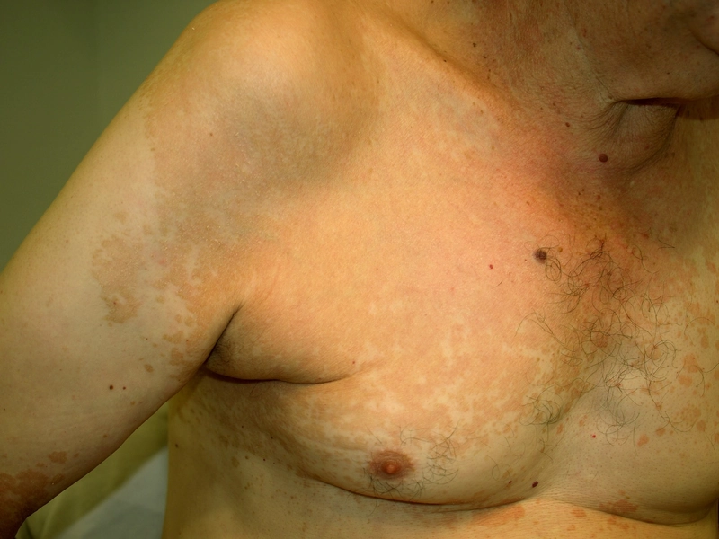
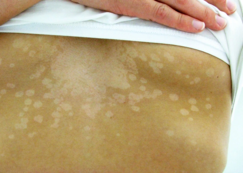
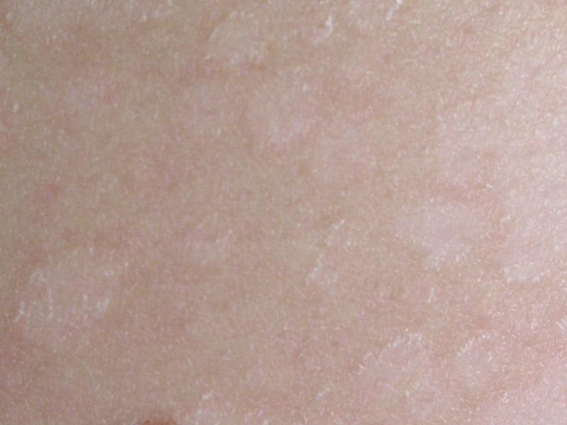
Definition
Malassezia (Yeasts)
- Affects: seborrheic areas (scalp, chest, upper back).
- Special form: Pityriasis versicolor — hypopigmentation due to tyrosinase inhibition.
- Further associations: seborrheic eczema, Malassezia folliculitis, sebopsoriasis.
Clinical features
Malassezia-associated Findings
- Pityriasis versicolor: hypo-/hyperpigmented scaling macules on the trunk (Malassezia)
- Malassezia folliculitis: monomorphic follicular papulopustules on upper back
- Is pityriasis versicolor contagious? No.
- Malassezia belongs to the lipophilic resident flora of sebum-rich skin and can be found in virtually every adult.
- Disease arises only when, given the right predisposition, the yeast converts into its mycelial (hyphal) form.
- The same fact explains the high tendency to recur: the organism remains part of the normal skin flora.
- Cause: inhibition of tyrosinase by dicarboxylic acids produced by Malassezia, among them azelaic acid.
- Repigmentation: needs time and light exposure.
Pityriasis versicolor alba
- Causative agent: Malassezia furfur / globosa — produces azelaic acid, which inhibits melanocytes.
- Localization: typically upper back, décolletage, shoulders (sebum-rich).
- KOH smear: "Spaghetti-and-meatballs" pattern (hyphae + spores).
Diagnosis
Wood's Lamp
- Microsporum canis (tinea capitis): yellow-green fluorescent — classic microsporia detection in children.
- Trichophyton schoenleinii (favus): blue-white fluorescent.
- Trichophyton tonsurans / T. verrucosum: no fluorescence — important pitfall, since these common tinea-capitis pathogens stay negative today.
- Pityriasis versicolor (Malassezia furfur): yellow-orange/greenish fluorescent.
Differential diagnoses
 Tinea corporis
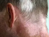 Vitiligo
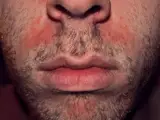 Seborrhoeic dermatitis
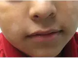 Pityriasis alba
 Hypomelanosis of Ito (incontinentia pigmenti achromians)
Image credits for the thumbnails
- Tinea corporis: Corina G., “Tinea corporis”, Public Domain, via Wikimedia Commons · Public domain · Source
- Vitiligo: Klaus D. Peter, Gummersbach, Germany, “Vitiligo and Poliosis”, CC BY 3.0 de · CC BY 3.0 de · Source
- Seborrhoeic dermatitis: Roymishali, “Seborrhoeic dermatitis highres”, CC BY-SA 3.0, via Wikimedia Commons · CC BY-SA 3.0 · Source
- Pityriasis alba: Siegfried EC, Hebert AA (J Clin Med 2015), CC BY 4.0, via PubMed Central (PMC4470205) · CC BY 4.0 · Source
- Hypomelanosis of Ito (incontinentia pigmenti achromians): Trägårdh M, Thomsen CR, Thorninger R et al. (J Med Case Rep 2014), “Hypomelanosis of Ito presenting with pediatric orthopedic issues: a case report”, CC BY 2.0, via PubMed Central (PMC4077238) · CC BY 2.0 · Source
Treatment principles
- Treatment is primarily topical, using antifungal shampoos, solutions or creams applied extensively to the affected areas.
- Systemic antifungals and intermittent topical prophylaxis against relapse are options for extensive disease or frequent recurrences.
- Hypopigmentation often persists for weeks to months after successful treatment and does not indicate treatment failure.
Specific treatment regimens with drugs and doses are covered in the DermaFuchs app.
Keep learning in the app
References (selection)
Cross-references
Mentioned in
Keep learning: test yourself
Two questions from the DermaFuchs app on Pityriasis versicolor (tinea versicolor) – tap an answer.
Question 1 of 2
To which ecological niche does the yeast Malassezia naturally belong in humans?
Answer
Correct: lipophilic resident flora of sebum-rich skin. Malassezia species are part of the normal lipophilic resident flora on sebum-rich areas of human skin.
Question 2 of 2
What is the classic microscopic appearance of Malassezia species in a direct smear?
Answer
Correct: spaghetti-and-meatballs phenomenon. Malassezia presents as a mix of short hyphae and round spores, often described as the 'spaghetti-and-meatballs' phenomenon.
More questions like these – with explanations and spaced repetition – in the app.
Note: This page is intended for medical education and does not replace diagnosis or treatment decisions in individual cases.