Naevus anaemicus (nevus anemicus)
- Synonyms
- Nävus anämicus, Nevus anemicus, pharmakologischer Nävus, pharmacological naevus
- Specialty
- Dermatology · Vascular skin disorders
- Images
- Clinical 1
- In the app
- 4 flashcards
- Last updated
- 10/2026 · Dr. Pascal Bafteh
Contents
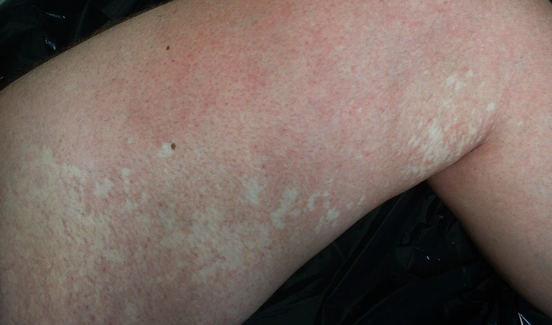
Images (1)
Definition
Naevus anaemicus is a congenital, localised vascular anomaly: a pale area of histologically normal skin whose pallor results from permanent vasoconstriction. Because of the altered vascular response it is also called a "pharmacological naevus".
Occurrence & epidemiology
It is usually noticed in early childhood but is probably present at birth; it occurs in all ethnic groups and is reported more often in females. It is frequent in neurofibromatosis type 1 (prevalence in children with NF1 8.8–51% depending on the study, higher at younger ages) and occurs in phakomatosis pigmentovascularis.
Aetiopathogenesis
The vessels within the lesion are hypersensitive to circulating catecholamines; via α-adrenergic receptors this leads to persistent vasoconstriction. In a transplantation study, the defect lay within the tissue of the naevus itself (donor dominance).
Clinical features
- Usually a single asymptomatic pale patch, round, oval or linear, often with small satellite macules; a mosaic form with multiple lesions appears net-like.
- Mainly on the trunk, in NF1 particularly on the anterior chest wall and often multiple; any body site possible.
- After rubbing, the patch does not redden, while the surrounding skin does.
- Occasionally other vascular anomalies nearby, e.g. a port-wine stain.
- Persists unchanged for life.
Diagnosis
- Clinical diagnosis, supported by the rubbing test.
- Diascopy (pressing the surrounding skin with a glass slide) eliminates the colour contrast.
- A biopsy shows normal skin.
- In children, the finding – especially when multiple – may point to NF1.
Differential diagnoses
Keep learning in the app
References (selection)
Cross-references
Note: This page is intended for medical education and does not replace diagnosis or treatment decisions in individual cases.