Progressive macular hypomelanosis
- Synonyms
- PMH
- Specialty
- Dermatology · Pigmentation disorders
- Images
- Clinical 1
- In the app
- 2 flashcards
- Last updated
- 10/2026 · Dr. Pascal Bafteh
Contents
Images (1)
Definition
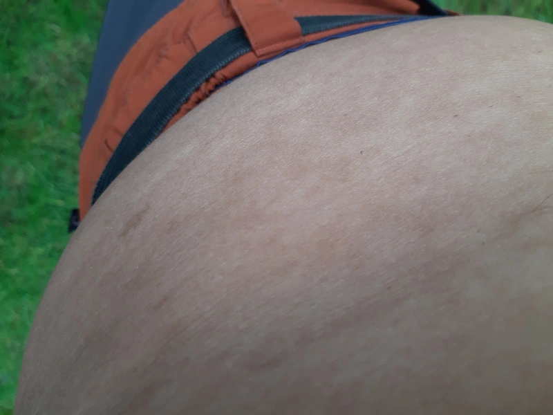
Progressive macular hypomelanosis is a common benign pigmentary disorder of teenagers and adults with ill-defined, non-scaly pale macules on the trunk that often coalesce. It is more frequent in darker skin types and is associated with Cutibacterium acnes in hair follicles.
- Causative agent: Cutibacterium acnes (old name Propionibacterium acnes).
- Localization: Trunk, especially lower back / generalized (in contrast to Pityriasis versicolor → upper back).
- Wood's light: red fluorescence (porphyrins).
Diagnosis
- Clinical; Wood's light shows follicular red fluorescence within the pale macules.
- Distinction mainly from hypopigmented pityriasis versicolor (scale, microscopy) and postinflammatory hypopigmentation; dermoscopy: poorly defined white macules with faint reticular pigmentation.
Differential diagnoses
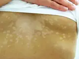 Pityriasis versicolor (tinea versicolor)
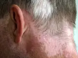 Vitiligo
 Naevus anaemicus (nevus anemicus)
 Pityriasis alba
 Idiopathic guttate hypomelanosis
Image credits for the thumbnails
- Pityriasis versicolor (tinea versicolor): Sarahrosenau on Flickr.com, “Tinea versicolor1”, CC BY-SA 2.0, via Wikimedia Commons · CC BY-SA 2.0 · Source
- Vitiligo: Klaus D. Peter, Gummersbach, Germany, “Vitiligo and Poliosis”, CC BY 3.0 de · CC BY 3.0 de · Source
- Naevus anaemicus (nevus anemicus): Birthmarkshare, “Nevus anemicus”, Public Domain, via Wikimedia Commons · Public domain · Source
- Pityriasis alba: Siegfried EC, Hebert AA (J Clin Med 2015), CC BY 4.0, via PubMed Central (PMC4470205) · CC BY 4.0 · Source
- Idiopathic guttate hypomelanosis: Kim Y, Shin K, Kim HS et al. (JAAD Int 2026), CC BY 4.0, via PubMed Central (PMC13524704) · CC BY 4.0 · Source · cropped
Treatment principles
- There is no established standard treatment; the condition is harmless and may regress spontaneously.
- Topical antibacterial treatment and phototherapy are used, with variable success.
- Recurrence after stopping treatment is common.
Specific treatment regimens with drugs and doses are covered in the DermaFuchs app.
Keep learning in the app
References (selection)
Cross-references
Mentioned in
Keep learning: test yourself
Two questions from the DermaFuchs app on Progressive macular hypomelanosis – tap an answer.
Question 1 of 2
What causes the red fluorescence under Wood's light in progressive macular hypomelanosis?
Answer
Correct: porphyrins. Porphyrins produced by Cutibacterium acnes cause the red fluorescence observed under Wood's light in progressive macular hypomelanosis.
Question 2 of 2
What is the possible cause of progressive macular hypomelanosis mainly on the trunk?
Answer
Correct: Propionibacterium acnes (Cutibacterium). Progressive macular hypomelanosis is possibly caused by Propionibacterium acnes (Cutibacterium).
More questions like these – with explanations and spaced repetition – in the app.
Note: This page is intended for medical education and does not replace diagnosis or treatment decisions in individual cases.