Idiopathic guttate hypomelanosis
- Synonyms
- Hypomelanosis guttata idiopathica, IGH, weiße Altersflecken, Leukopathia guttata, white sunspots
- Specialty
- Dermatology · Pigmentation disorders
- Images
- Clinical 1
- In the app
- 2 flashcards
- Last updated
- 10/2026 · Dr. Pascal Bafteh
Contents
Images (1)
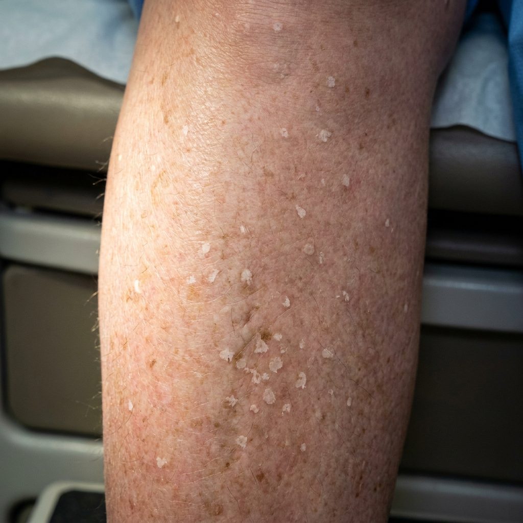 AI illustration
Definition
Idiopathic guttate hypomelanosis is a common, benign, acquired leukoderma with small, well-defined, porcelain-white macules on sun-exposed limbs.
Occurrence & epidemiology
Both sexes and all skin types are affected. It mostly affects people over 40 years, and its frequency rises with age – up to about 90% in the ninth decade. It is uncommon in children and teenagers; familial cases are common.
Aetiopathogenesis
Probably multifactorial: skin ageing, chronic UV exposure, genetic factors, trauma and autoimmunity. Abnormal uptake of melanin by keratinocytes has been demonstrated, reducing pigment transfer from melanocytes.
Clinical features
- Smooth, hypo- or depigmented, round to oval macules of 2–5 mm (up to 1.5 cm), usually multiple and asymptomatic.
- Mainly on the extensor forearms, the shins and the V of the chest.
- More obvious in skin of colour.
- Variants: hypopigmented macules on sun-damaged skin; a single white stellate sclerotic macule; small macules with a scalloped margin and hyperkeratotic surface.
Histology
Flattened, thin epidermis with basket-weave hyperkeratosis and loss of rete ridges; melanin in the epidermis reduced or absent; melanocytes normal or reduced, with short retracted dendrites; small foci of retained melanin in the basal layer.
Diagnosis
- Usually a clinical diagnosis.
- Wood lamp emphasises the loss of pigment.
- Dermoscopy: well-defined white structureless areas, absent pigment network.
- Biopsy only to exclude other leukodermas, such as vitiligo or lichen sclerosus.
Differential diagnoses
Keep learning in the app
References (selection)
Cross-references
Mentioned in
Note: This page is intended for medical education and does not replace diagnosis or treatment decisions in individual cases.