Purpura fulminans
- Synonyms
- akut-infektiöse Purpura fulminans, Purpura fulminans neonatalis, postinfektiöse Purpura fulminans, acute infectious purpura fulminans, neonatal purpura fulminans, idiopathic purpura fulminans
- Specialty
- Dermatology · Vascular skin disorders
- Images
- Clinical 1
- In the app
- 2 flashcards
- Last updated
- 10/2026 · Dr. Pascal Bafteh
Contents
Images (1)
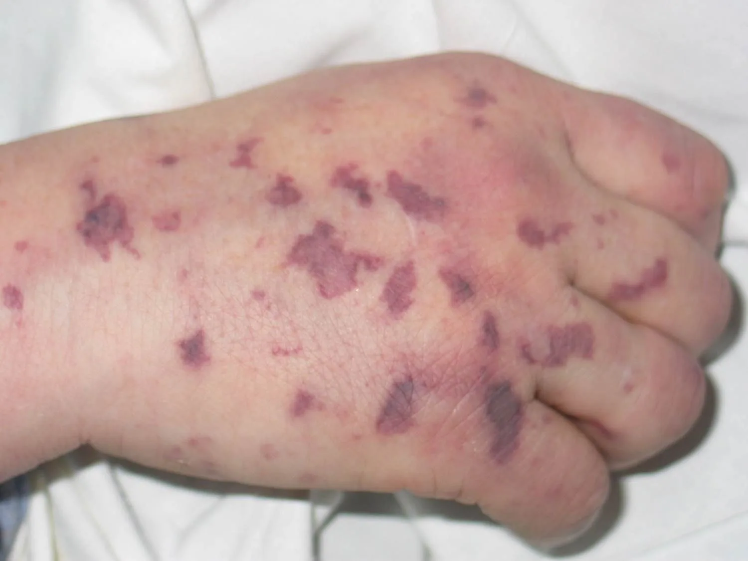
Definition
Purpura fulminans is a rare, rapidly progressive and often fatal, highly thrombotic form of disseminated intravascular coagulation (DIC). Thrombotic occlusion of small and medium-sized vessels leads to skin necrosis, shock and multi-organ failure – a dermatological and haematological emergency.
Classification
- Neonatal form: fewer than 1 in 1,000,000 births; severe congenital protein C or protein S deficiency; onset hours to days after birth.
- Acute infectious form (most common): in severe infections and sepsis, including meningococci, streptococci, Staphylococcus aureus, Haemophilus and Clostridia.
- Idiopathic (post-infectious) form: 7–10 days after an infection (especially group A streptococcus, varicella) due to autoantibodies against protein S.
Occurrence & epidemiology
Children are most often affected, with two peaks at 1–3 and 16–18 years of age; adults can also be affected.
Aetiopathogenesis
The basis is a shift of haemostasis towards coagulation; in the infectious form, impaired protein C function or acquired protein C deficiency, among other factors, is implicated. Microvascular thrombosis causes the skin necrosis.
Clinical features
- Onset with skin pain, erythematous macules and petechiae, followed by ecchymoses and painful indurated purpuric plaques with erythematous borders.
- Classically retiform purpura with branched or angular lesions, particularly on the extremities.
- Within 24–48 hours haemorrhagic blisters and full-thickness skin necrosis.
- Systemically sepsis with or without shock, sometimes meningeal signs. Mortality of meningococcal disease complicated by purpura fulminans is 20–60%; survivors often have amputations and extensive scarring.
Diagnosis
- Clinical (acute, widespread retiform purpura); urgent skin biopsy to exclude other causes; blood or tissue cultures to identify the organism.
- Laboratory: thrombocytopenia, prolonged clotting times, decreased fibrinogen, elevated D-dimer; measurement of protein C, protein S and antithrombin III.
Differential diagnoses
Keep learning in the app
References (selection)
Cross-references
Mentioned in
Note: This page is intended for medical education and does not replace diagnosis or treatment decisions in individual cases.