Warfarin-induced skin necrosis (coumarin necrosis)
- Synonyms
- Cumarinnekrose, Warfarin-Nekrose, Phenprocoumon-Nekrose, Coumarin necrosis, Warfarin necrosis, Warfarin-induced skin necrosis
- Specialty
- Dermatology · Drug reactions
- Images
- Clinical 1
- In the app
- 1 flashcards
- Last updated
- 10/2026 · Dr. Pascal Bafteh
Contents
Images (1)
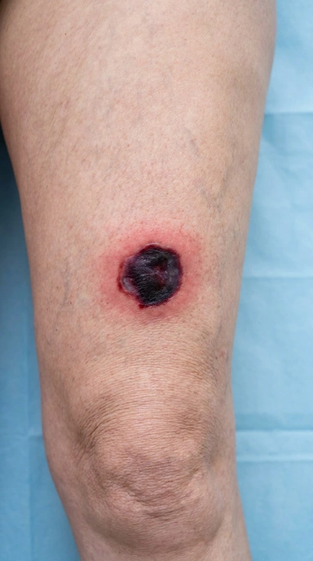 AI illustration
Definition
Coumarin necrosis is a rare, severe complication induced by coumarin-type vitamin K antagonists (e.g. warfarin, phenprocoumon): paradoxical clot formation in small skin vessels causes necrosis of the skin and subcutaneous fat. It was first described in 1943.
Occurrence & epidemiology
After taking warfarin, it occurs in about 1 in 10,000 people (reported prevalence 0.01–0.1%). Women are affected more often, usually between 50 and 70 years of age; obesity increases the risk.
Aetiopathogenesis
Vitamin K antagonists lower clotting factors II, VII, IX and X but also the natural clotting regulators protein C and protein S. Protein C has a half-life of about 6 hours, factors II and X of 2–5 days; in the first days after starting the drug this creates a transient prothrombotic state. Predisposing factors are inherited protein C or protein S deficiency, factor V Leiden, antithrombin III deficiency and antiphospholipid antibodies.
Clinical features
- Onset usually between day 2 and day 5 after starting the drug, very rarely after weeks or months.
- First pain and purpura, which turns bluish-black with a red rim within days; then haemorrhagic blisters and full-thickness skin necrosis, sometimes with retiform purpura.
- Predilection for fatty areas: breasts, thighs, buttocks, hips, abdomen.
Histology
Diffuse microthrombi in dermal vessels with endothelial damage and red cell extravasation, without significant inflammation; later full-thickness coagulative necrosis.
Diagnosis
- Clinical diagnosis from timing and distribution.
- Skin biopsy for support and to distinguish it from necrotising fasciitis and calciphylaxis, among others.
- Protein C and protein S blood levels to look for a predisposition.
Differential diagnoses
Keep learning in the app
References (selection)
Cross-references
Note: This page is intended for medical education and does not replace diagnosis or treatment decisions in individual cases.