Staphylococcal scalded skin syndrome (SSSS)
- Synonyms
- SSSS, staphylogenes Lyell-Syndrom, Morbus Ritter, Staphylococcal scalded skin syndrome, Ritter disease
- Specialty
- Dermatology · Bacterial infections
- Images
- Clinical 1
- In the app
- 5 flashcards
- Last updated
- 10/2026 · Dr. Pascal Bafteh
Contents
Images (1)
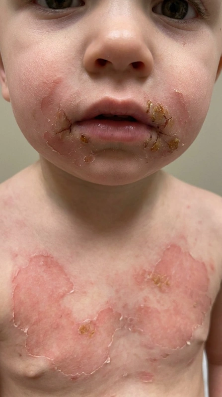 AI illustration
Definition
Staphylococcal scalded skin syndrome (SSSS) is a rare, severe, superficial blistering disorder triggered by exfoliative toxins from certain strains of Staphylococcus aureus. Widespread detachment of the uppermost layers of the epidermis makes the skin look scalded. In newborns the name Ritter disease is also used.
Occurrence & epidemiology
It mainly affects children younger than 5 years (peak at 2–3 years), explained by a lack of antibodies against the toxins and immature renal toxin clearance. Adults are rarely affected, usually with immunosuppression or renal impairment. For Europe, 0.09–0.56 cases per million people have been reported.
Aetiopathogenesis
The starting point is a localised infection with toxin-producing S. aureus (about 5% of strains), e.g. of the conjunctiva, throat, umbilicus or a wound. Exfoliative toxins A and B, spread via the bloodstream, cleave desmoglein 1 – hence the very superficial cleavage plane in the granular layer. Desmoglein 1 is absent from mucosal epithelium, so the mucous membranes are spared.
Clinical features
- Fever and irritability; within 24–48 hours a painful, rapidly spreading erythema, starting on the face and in the flexures.
- The skin is exquisitely tender with a wrinkled, tissue-paper-like consistency; large, flaccid blisters at sites of friction. The epidermis peels off in sheets, leaving moist, scalded-looking areas.
- Nikolsky sign I positive: gentle rubbing of normal-looking skin shears off the epidermis.
- Mucous membranes spared – an important difference from toxic epidermal necrolysis (TEN).
Histology
Non-inflammatory, superficial splitting within the granular layer. In TEN, by contrast, the cleavage plane is subepidermal, at or below the basal cell layer.
Diagnosis
- Usually a clinical diagnosis.
- Cultures from the suspected source of infection (conjunctiva, nasopharynx, umbilicus); blister fluid is usually sterile.
- Biopsy with frozen section in unclear cases to determine the cleavage plane rapidly and distinguish it from TEN.
Differential diagnoses
- Stevens-Johnson syndrome
- Impetigo (impetigo contagiosa)
- Staphylococcal scalded skin syndrome
- Necrolytic migratory erythema
- Pemphigus vulgaris
- Acanthosis nigricans maligna
Keep learning in the app
References (selection)
Cross-references
Mentioned in
Note: This page is intended for medical education and does not replace diagnosis or treatment decisions in individual cases.