Frostbite
- Synonyms
- Congelatio, Frostschaden, Kälteschaden, frostnip, cold injury
- Specialty
- Dermatology · Environmental & photodermatoses
- Images
- Clinical 4
- In the app
- 1 flashcard
- Last updated
- 10/2026 · Dr. Pascal Bafteh
Contents
Images (4)
Definition
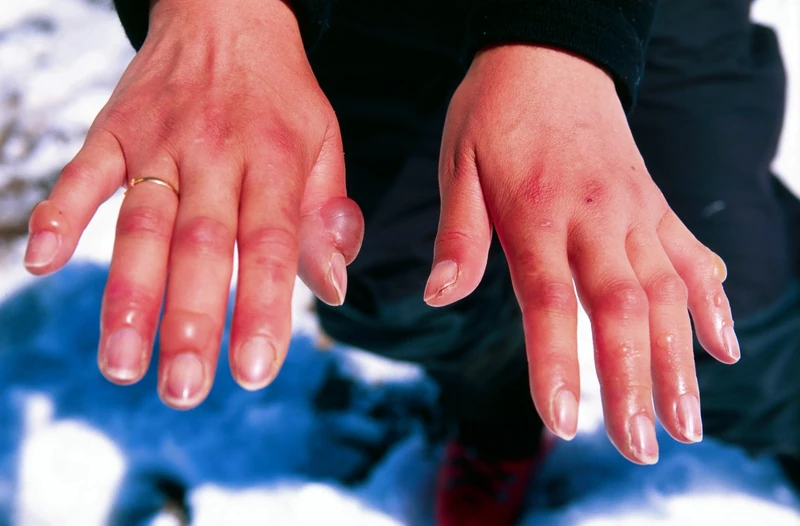
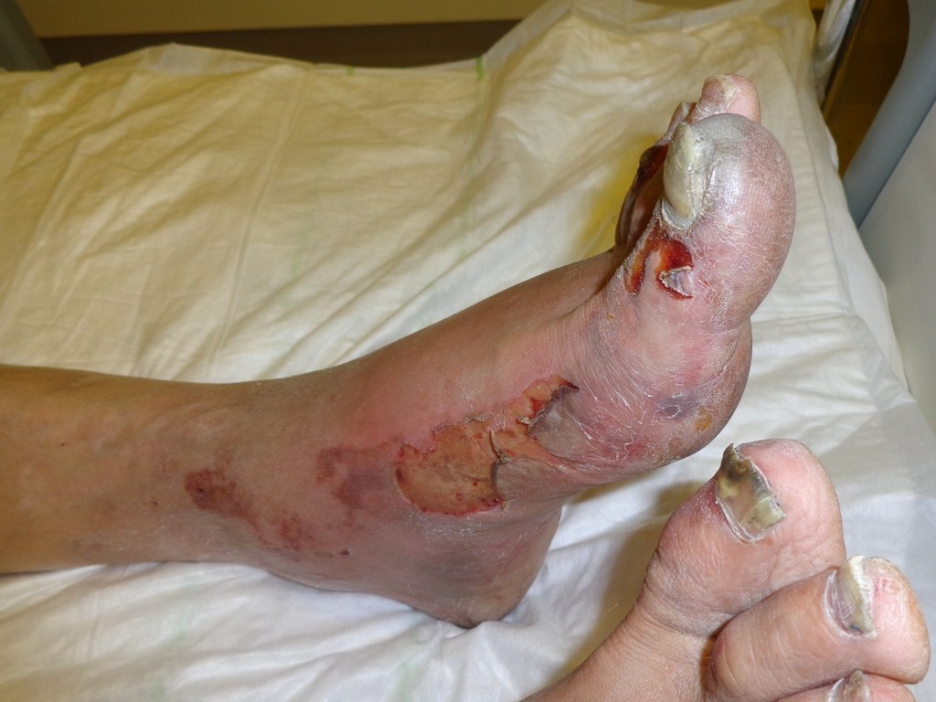
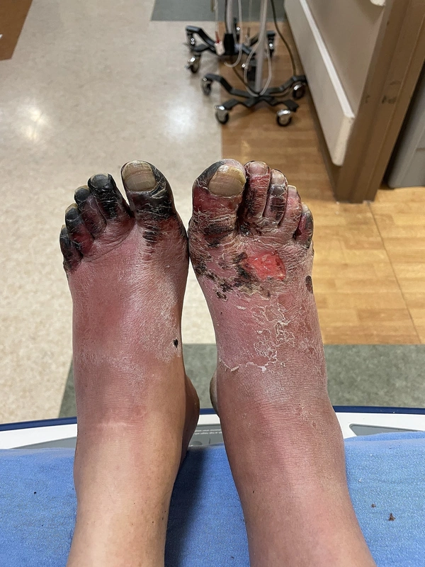
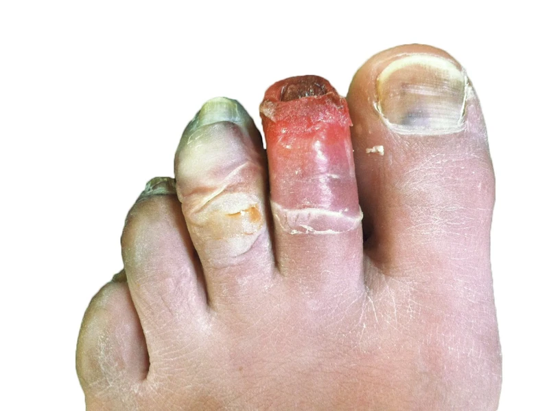
Frostbite is local tissue injury due to cold below freezing. Ice crystal formation in the tissue, cell damage and disturbed microcirculation with thrombosis lead to tissue loss, which often becomes fully demarcated only after days.
Classification
- Grade 1 (frost nip): involves only the top layer of skin (epidermis).
- Grade 2 (superficial frostbite): white or blue skin that feels hard and frozen; blisters with clear or milky fluid form within 24 hours, the underlying tissue is still intact.
- Grade 3 (deep frostbite): white, blotchy or blue skin, the underlying tissue is damaged; blood-filled blisters that form thick black scabs.
- Grade 4: full-thickness damage affecting muscles, tendons and bone, with tissue loss.
Occurrence & epidemiology
Those at risk include mountaineers and winter athletes, people stranded or working in the cold (e.g. soldiers, rescuers), homeless people, the very young and the very old, people with peripheral vascular disease or diabetes mellitus, and smokers or people taking vasoconstricting drugs (e.g. beta blockers). Mainly the toes, feet, fingers, nose, ears and cheeks are affected.
Diagnosis
- Clinical grading after rewarming; the final extent is often visible only after days to weeks.
- Imaging (scintigraphy, MR angiography) to assess perfusion in severe frostbite.
Differential diagnoses
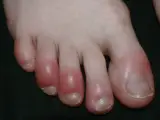 Pernio (Chilblain)
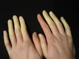 Raynaud syndrome
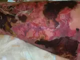 Necrotizing fasciitis
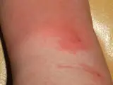 Erysipelas
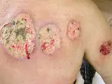 Pyoderma gangrenosum
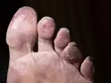 Cholesterol embolism (atheroembolism)
 Warfarin-induced skin necrosis (coumarin necrosis)
Image credits for the thumbnails
- Pernio (Chilblain): Sapp, Public domain, via Wikimedia Commons · Public Domain · Source
- Raynaud syndrome: Niklas D, “Raynaud-Syndrom”, CC BY-SA 3.0, via Wikimedia Commons · CC BY-SA 3.0 · Source
- Necrotizing fasciitis: CNX OpenStax, CC BY 4.0, via Wikimedia Commons, cropped · CC BY 4.0 · Source
- Erysipelas: Poupou l'quourouce, “Erysipel”, CC BY-SA 3.0, via Wikimedia Commons · CC BY-SA 3.0 · Source
- Pyoderma gangrenosum: Monopol, “Pyoderma gangrenosum 01”, CC BY-SA 3.0, via Wikimedia Commons · CC BY-SA 3.0 · Source
- Cholesterol embolism (atheroembolism): Akpala C, Kim G, Froehling D et al. (Cureus 2023), CC BY 4.0, via PubMed Central (PMC10640895) · CC BY 4.0 · Source
- Warfarin-induced skin necrosis (coumarin necrosis): Zahra T, Jamil S, Ferman H et al. (Cureus 2022), CC BY 3.0, via PubMed Central (PMC9664942) · CC BY 3.0 · Source
Treatment principles
- Concomitant hypothermia is treated first; local initial treatment consists of rapid rewarming in a warm water bath once refreezing can be excluded, with adequate analgesia and without rubbing the skin.
- This is followed by stage-appropriate wound care and checking of tetanus immunisation.
- Surgery is deferred until the tissue has clearly demarcated; in severe, recent frostbite, thrombolysis may be considered in specialised centres.
Specific treatment regimens with drugs and doses are covered in the DermaFuchs app.
Keep learning in the app
References (selection)
Cross-references
Keep learning
Keep learning with flashcards, exam questions and spot diagnoses in the DermaFuchs app – free, in your browser or as an app.
Related pages: Pernio (Chilblain) · Raynaud syndrome · Necrotizing fasciitis
Note: This page is intended for medical education and does not replace diagnosis or treatment decisions in individual cases.