Syringoma
- Synonyms
- Syringome, eruptive Syringome, Syringom Augenlid, syringomas, eruptive syringoma
- Specialty
- Dermatology · Skin tumours
- Images
- Clinical 1 · Histology 1
- In the app
- 2 flashcards
- Last updated
- 10/2026 · Dr. Pascal Bafteh
Contents
Images (2)
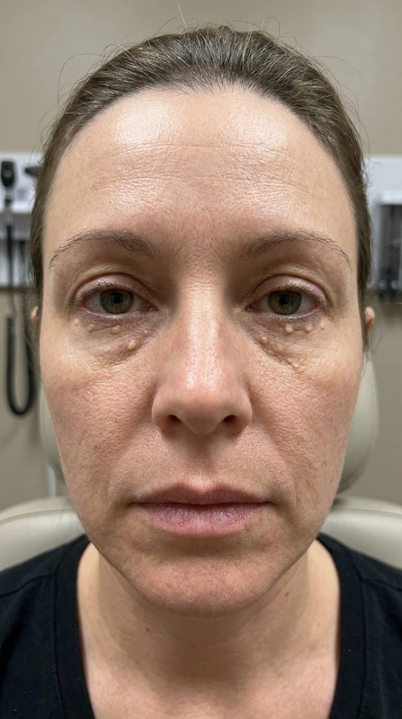 AI illustration
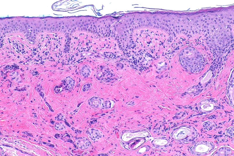 Histology
Definition
A syringoma is a benign adnexal tumour arising from the acrosyringium, the intraepidermal portion of the eccrine sweat duct. Syringomas typically occur in clusters on and around the eyelids.
Classification
Friedman and Butler distinguish:
- localised syringomas
- generalised multifocal or eruptive syringomas
- syringomas associated with Down syndrome
- familial syringomas
The clear cell syringoma, with glycogen deposition and an association with diabetes mellitus, is rare.
Occurrence & epidemiology
Syringomas affect about 1% of the population, women twice as often as men. They are usually sporadic and appear during or after puberty. Familial syringomas are inherited in an autosomal dominant manner and often begin before puberty. In trisomy 21, 18–39% of individuals have syringomas.
Clinical features
- Firm, round, skin-coloured dermal papules of 1–3 mm, solitary or clustered, often symmetrical.
- Predilection: eyelids and cheeks; also scalp, axillae, umbilicus, chest and genital region.
- Eruptive syringomas: crops of yellow-brown to hyperpigmented papules on the neck, anterior trunk, axillae, shoulders and abdomen.
- Usually asymptomatic; itching may occur with sweating or on the vulva.
Histology
Normal epidermis; in the dermis numerous small ducts and epithelial cords in a sclerotic stroma. The ducts are lined by two rows of flattened epithelial cells; the outer layer bulges outwards into a comma-like tail (tadpole shape). In clear cell syringoma the cytoplasm is pale owing to glycogen.
Diagnosis
- Usually a clinical diagnosis based on the typical appearance.
- A full-thickness skin biopsy is needed to distinguish microcystic adnexal carcinoma, which infiltrates the deep dermis and subcutis.
- Eruptive syringomas are often not suspected clinically and are only recognised histologically.
- In clear cell syringoma, assessment for impaired glucose tolerance is indicated.
Differential diagnoses
- Trichoepithelioma
- Hidrocystoma (eccrine and apocrine)
- Milia (milium)
- Basal cell carcinoma
- Fibrofolliculoma (Birt-Hogg-Dubé)
- Epidermoid cyst
- Sebaceous hyperplasia
Keep learning in the app
References (selection)
Cross-references
Note: This page is intended for medical education and does not replace diagnosis or treatment decisions in individual cases.