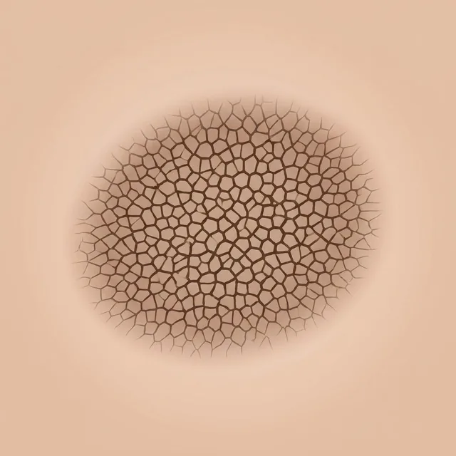
 AI schematic
Network
A fine, regular grid of brown lines with lighter holes. The lines correspond to the pigmented rete ridges.
Typical of: Melanocytic naevus, Dysplastic naevus
Also in German (Deutsch)
84 structures and patterns from the four learning cards by Ralph P. Braun and Ashfaq A. Marghoob – some with a schematic, each with one sentence on what it means and the diagnoses in which it is typical.
About the images: All images are idealised schematic illustrations (partly AI-generated, partly drawn programmatically) and do not replace a photograph. Each image is labelled accordingly. Names and outline follow the Braun/Marghoob learning cards; the card images themselves are not shown here.
AI schematicA fine, regular grid of brown lines with lighter holes. The lines correspond to the pigmented rete ridges.
Typical of: Melanocytic naevus, Dysplastic naevus
Pigment network present only in islands, with structureless or homogeneous areas in between.
Typical of: Melanocytic naevus, Dysplastic naevus
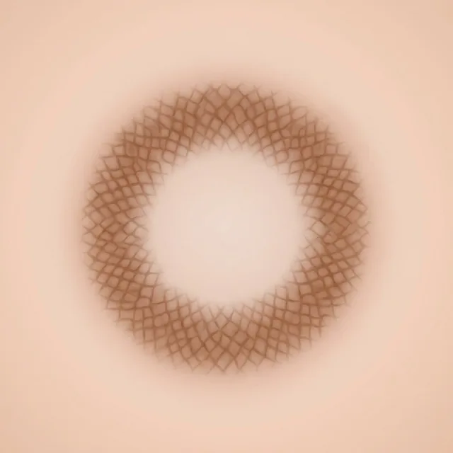 AI schematic
Network at the periphery, a lighter structureless zone in the centre.
Typical of: Melanocytic naevus, Dysplastic naevus
Network at the periphery, a darker structureless zone in the centre. A central dark zone fits a naevus; an eccentric one is suspicious.
Typical of: Melanocytic naevus, Dysplastic naevus
Network at the periphery, brown globules in the centre.
Typical of: Melanocytic naevus
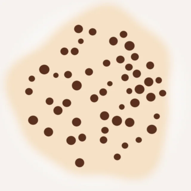 Schematic
Many round to oval brown globules of similar size and colour, evenly distributed. They correspond to nests of naevus cells.
Typical of: Melanocytic naevus
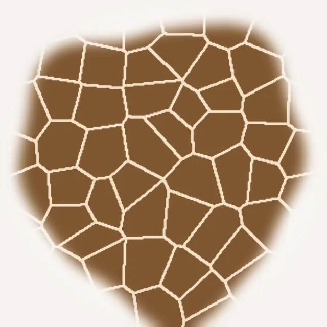 Schematic
Densely packed, angular, flattened globules resembling cobblestones.
Typical of: Melanocytic naevus
 AI schematic
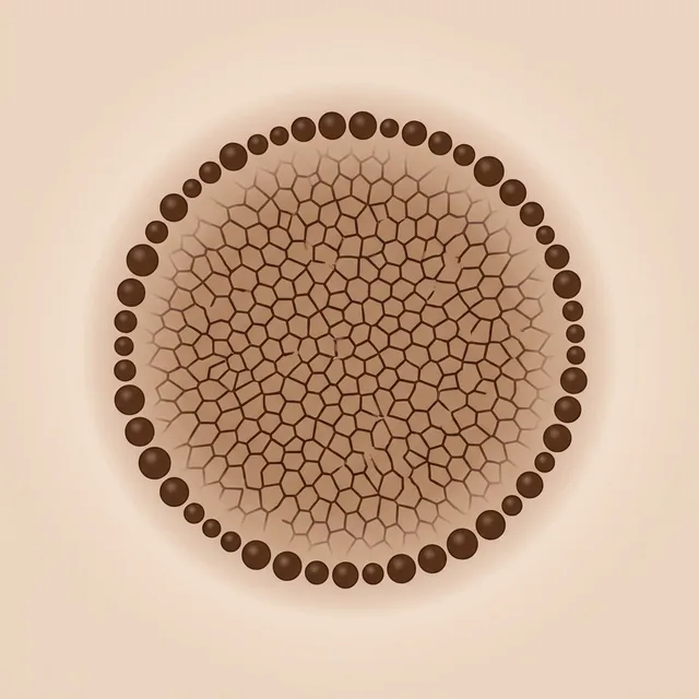
Network in the centre with a rim of brown globules at the edge – a sign of a growing naevus, mainly in younger people.
Typical of: Melanocytic naevus, Dysplastic naevus
 Schematic
Uniform steel-blue to blue-grey area without network or globules. The pigment lies deep in the dermis and therefore looks blue (Tyndall effect).
Typical of: Blue naevus
 Schematic
Uniform brown area without recognisable internal structures.
Typical of: Melanocytic naevus
 AI schematic
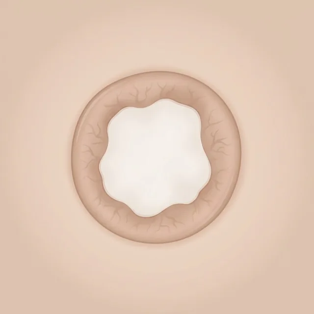
A delicate brown network at the periphery around a central white, scar-like patch.
Typical of: Dermatofibroma
Ring-like globules (light centre, brown rim), shiny white streaks under polarised light and a pink background.
Typical of: Dermatofibroma
A soft, skin-coloured to brownish papule with short comma-shaped vessels, often with a brown halo, brown pigmentation and globules.
Typical of: Melanocytic naevus
 Schematic
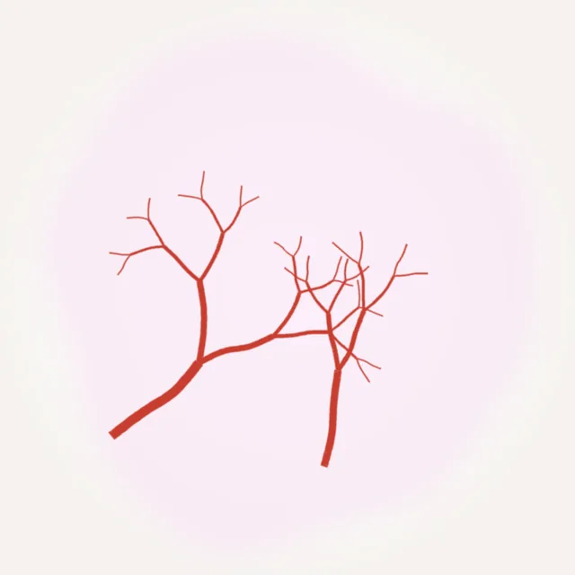
Sharply focused, tree-like branching red vessels that taper into fine branches.
Typical of: Basal cell carcinoma
Radial projections around a darker centre, like the spokes of a wheel.
Typical of: Basal cell carcinoma
Brown to blue-grey, leaf-like lobulated areas without a network, usually at the edge of the lesion.
Typical of: Basal cell carcinoma
Larger, well-defined oval blue-grey areas – pigmented tumour nests in the dermis.
Typical of: Basal cell carcinoma
 Schematic
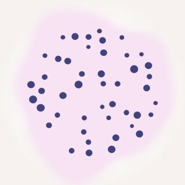
Many small, round blue-grey dots and globules that are not part of a network.
Typical of: Basal cell carcinoma
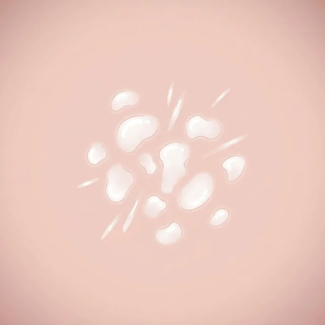 AI schematic
Shiny white structureless patches and short strands, visible under polarised light.
Typical of: Basal cell carcinoma
A structureless red to brown-black area, often crusted, where the epidermis is missing – suspicious if there was no preceding injury.
Typical of: Basal cell carcinoma, Squamous cell carcinoma
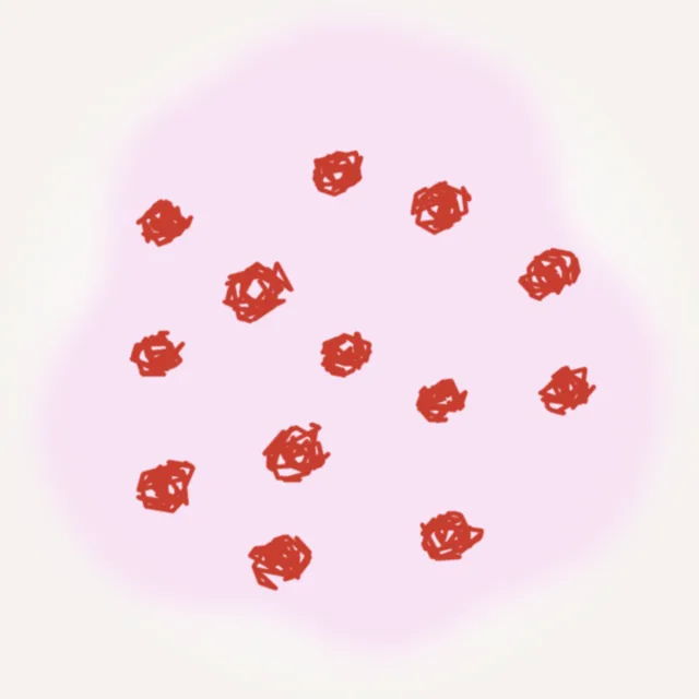 Schematic
Small, tightly coiled vessels, often in clusters – they resemble renal glomeruli.
Typical of: Bowen's disease, Squamous cell carcinoma
White rings around follicular openings.
Typical of: Squamous cell carcinoma, Keratoacanthoma
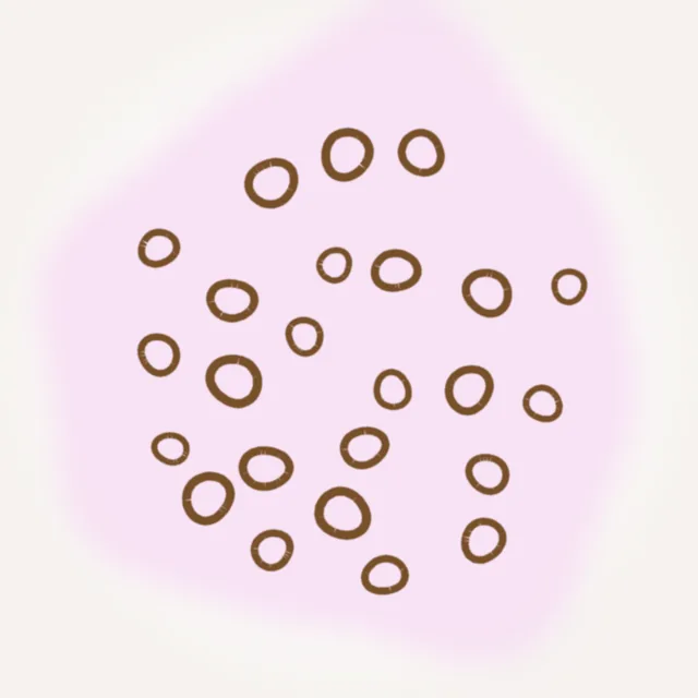 Schematic
Brown rings surrounding follicular openings. On the Braun/Marghoob cards they are grouped with the pigmented forms of the SCC spectrum; poorly studied and hard to distinguish from lentigo maligna (annular-granular).
Typical of: Actinic keratosis, Bowen's disease
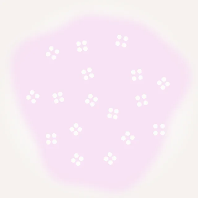 Schematic
Four white dots arranged like a four-leaf clover – visible only under polarised light. Non-specific, mainly AK/SCC.
Typical of: Actinic keratosis, Squamous cell carcinoma
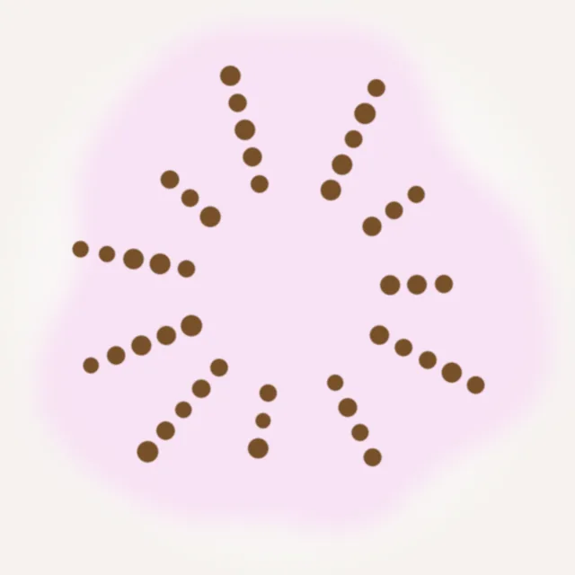 Schematic
Small brown dots lined up in rows, often radiating towards the edge.
Typical of: Bowen's disease
Yellowish structureless keratin on the surface.
Typical of: Actinic keratosis, Bowen's disease, Squamous cell carcinoma
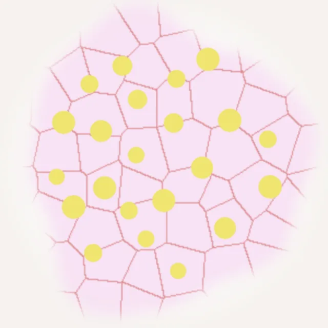 Schematic
A red pseudonetwork between the follicles with yellowish-white keratin-plugged follicular openings – like the surface of a strawberry, mainly on the face.
Typical of: Actinic keratosis
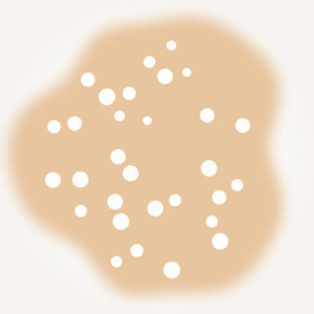 Schematic
Round white to yellowish dots, often shining like stars under non-polarised light – small horn cysts in the epidermis.
Typical of: Seborrhoeic keratosis
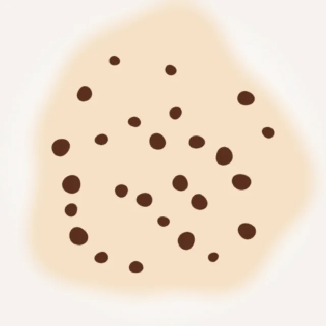 Schematic
Round to irregular brown to black plugs – keratin-filled invaginations of the surface.
Typical of: Seborrhoeic keratosis
Fine, parallel, curved light-brown lines that do not form a network – like a fingerprint.
Typical of: Solar lentigo, Seborrhoeic keratosis
 Schematic
A sharp, scalloped border that looks as if moths had nibbled at it.
Typical of: Solar lentigo, Seborrhoeic keratosis
 Schematic
An abrupt transition between lesion and surrounding skin, with no gradual fading.
Typical of: Seborrhoeic keratosis
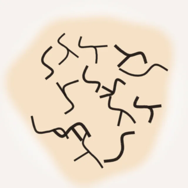 Schematic
Raised ridges (gyri) with darker furrows (sulci) in between – reminiscent of the surface of a brain.
Typical of: Seborrhoeic keratosis
Hairpin-shaped vascular loops, each surrounded by a whitish halo.
Typical of: Seborrhoeic keratosis, Keratoacanthoma
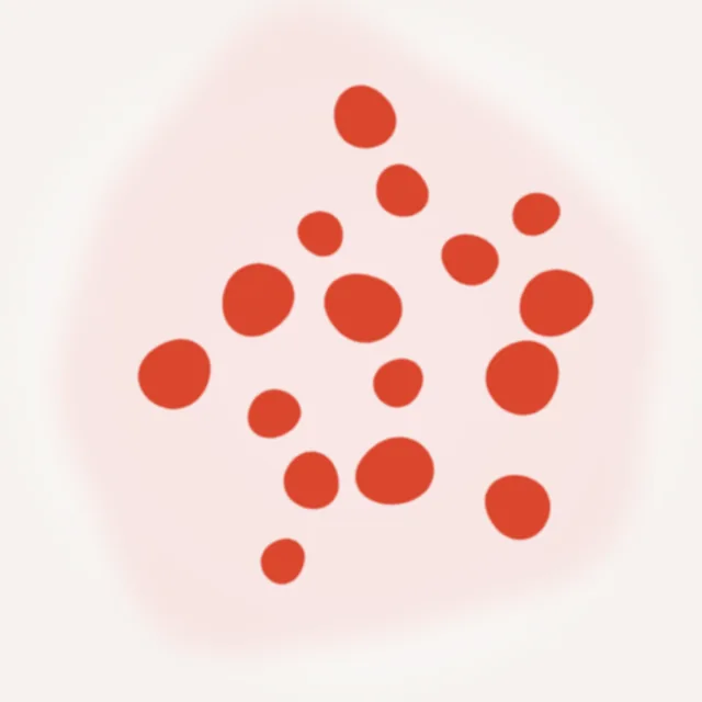 Schematic
Sharply demarcated round to oval red areas (lacunae), separated by pale septa – dilated vascular spaces.
Typical of: Angioma
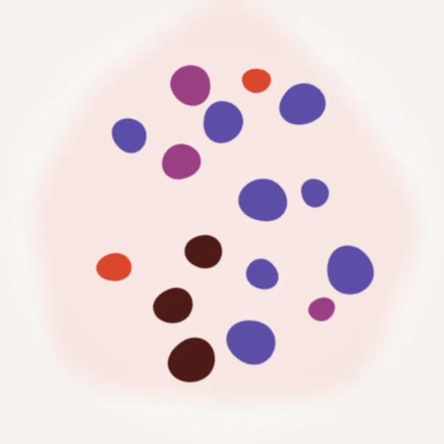 Schematic
Lacunae in red, blue-violet to black (thrombosed), often with a whitish veil or crust.
Typical of: Angiokeratoma
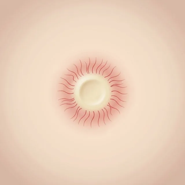 AI schematic
Yellowish-white lobules with vessels that run from the edge towards the centre without crossing it.
Typical of: Sebaceous hyperplasia
Dotted or coiled vessels lined up in serpiginous rows like a string of pearls.
Typical of: Clear cell acanthoma
Pigment on the ridges rather than in the furrows; the sweat-duct openings show as white dots within the pigment.
Typical of: Melanoma
Fine pigment lines running obliquely across the ridges which – unlike the even fibrillar pattern of a naevus – vary in colour, thickness and density.
Typical of: Melanoma
 AI schematic
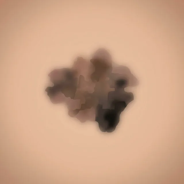
Irregular, diffuse pigmentation in several shades of brown to grey without any recognisable order.
Typical of: Melanoma
Three or more different patterns within one acral lesion.
Typical of: Melanoma
Follicular openings with uneven grey-brown pigmentation, often on one side only.
Typical of: Lentigo maligna
Fine grey to brown dots arranged in rings around the follicular openings.
Typical of: Lentigo maligna
Thickened grey-brown lines that surround the follicular openings in a rhomboid shape.
Typical of: Lentigo maligna
Dark structureless areas in which the follicular openings disappear.
Typical of: Lentigo maligna
Red-purple to black-brown patches or globules beneath the nail plate, often sharply demarcated; the discolouration grows out with the nail.
Typical of: Subungual haematoma
Fungal infection: yellowish-white streaks with jagged, spike-like projections pointing proximally. Pseudomonas: greenish discolouration of the nail plate.
Typical of: Onychomycosis, Green nail syndrome
 Schematic
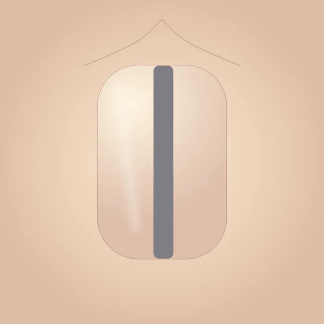
A grey longitudinal band with grey lines: the melanocytes of the nail matrix produce more pigment without increasing in number.
Typical of: Longitudinal melanonychia (e.g. ethnic, lentigo)
A brown longitudinal band: a sign of an increased number of melanocytes in the nail matrix. Whether it is regular (naevus) or irregular (melanoma) is shown by the lines within the band.
Typical of: Melanocytic naevus, Melanoma
 AI schematic
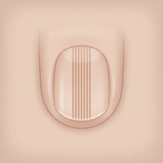
Brown longitudinal lines of equal width, spacing and colour, parallel throughout.
Typical of: Melanocytic naevus
 AI schematic
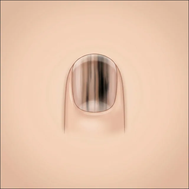
Brown to black longitudinal lines varying in width, colour and spacing and losing their parallelism. Pigment on the nail fold (Hutchinson sign) is an additional warning sign.
Typical of: Melanoma
Two different patterns within one lesion, such as network and globules. Possible in naevi – interpretation needs age, site and history.
Typical of: Melanocytic naevus
Tan to pink and structureless, without network or globules. Often a naevus in very fair skin, but only interpretable in context because amelanotic melanomas can look similar.
Typical of: Melanocytic naevus, Amelanotic melanoma
Three or more patterns within one lesion, but arranged symmetrically.
Typical of: Melanocytic naevus, Dysplastic naevus
Globules in several rows around the whole lesion – a sign of symmetrical outward growth. New onset at an older age warrants excluding melanoma.
Typical of: Spitz/Reed naevus
Streaks or pseudopods radiating evenly all around a dark centre. New onset at an older age warrants excluding melanoma.
Typical of: Spitz/Reed naevus
Three or more different patterns, distributed asymmetrically.
Typical of: Melanoma
The whole lesion is predominantly structureless brown, without network or globules. A feature-poor melanoma can look like this, especially when no benign naevus pattern is present.
Typical of: Melanoma
A pink to tan, structureless flat lesion; this is how an amelanotic or hypomelanotic melanoma can present.
Typical of: Amelanotic melanoma, Melanoma
Small, irregularly distributed darker islands or foci at the edge of an otherwise pale lesion.
Typical of: Melanoma
Tan structureless areas interspersed with fine grey dots (granularity).
Typical of: Melanoma, Lentigo maligna
Straight, angulated lines that may join to form polygons – suspicious of melanoma on sun-damaged skin.
Typical of: Melanoma, Lentigo maligna
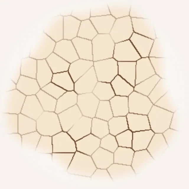 Schematic
Pigment network with lines of uneven thickness, partly dark, and irregular holes, unevenly distributed across the lesion.
Typical of: Melanoma, Dysplastic naevus
Straight lines meeting at angles and forming angular figures instead of round network holes.
Typical of: Melanoma, Lentigo maligna
Light serpiginous lines enclosing darker, elongated globules in a net-like fashion – the negative of a pigment network.
Typical of: Melanoma, Spitz/Reed naevus
Streaks or pseudopods (streaks with a bulbous tip) found only on part of the edge or distributed unevenly.
Typical of: Melanoma
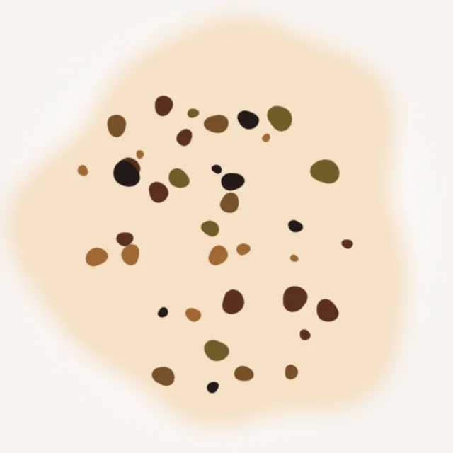 Schematic
Dots and globules varying in size, colour and shape, distributed unevenly.
Typical of: Melanoma, Dysplastic naevus
Tan areas without structures at the periphery of an otherwise structured lesion.
Typical of: Melanoma
The skin furrows stand out much more within the lesion than in the surrounding skin – together with small foci of hyperpigmentation a clue to melanoma in situ.
Typical of: Melanoma
Several small, ill-defined darker spots within one lesion.
Typical of: Melanoma
Dark structureless blotches, irregular in shape and position, often eccentric.
Typical of: Melanoma
An ill-defined blue area with a whitish, ground-glass veil over a raised part of the lesion.
Typical of: Melanoma
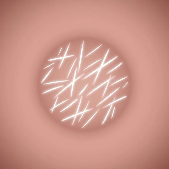 AI schematic
Shiny white short lines lying parallel or at right angles to each other, or shiny white patches, visible under polarised light.
Typical of: Melanoma, Basal cell carcinoma, Spitz/Reed naevus, Dermatofibroma
Fine grey dots like ground pepper – melanin within melanophages in the upper dermis.
Typical of: Melanoma, Lentigo maligna, Lichenoid keratosis
Scar-like white areas, lighter than the surrounding skin, with fine grey dots.
Typical of: Melanoma
A flat blue-white structureless area that is not raised – a sign of regression rather than of a nodular component.
Typical of: Melanoma
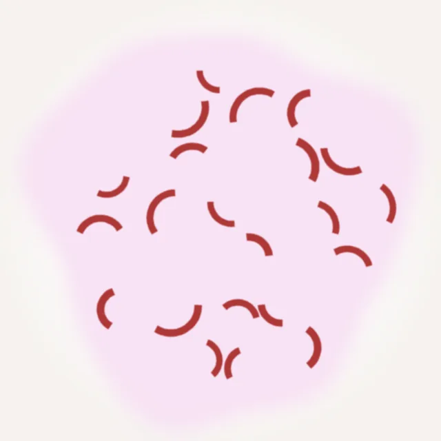 Schematic
Short curved vessels shaped like a comma. Typical of dermal naevi; in a flat lesion alongside other vessel types a clue to melanoma.
Typical of: Melanocytic naevus, Melanoma
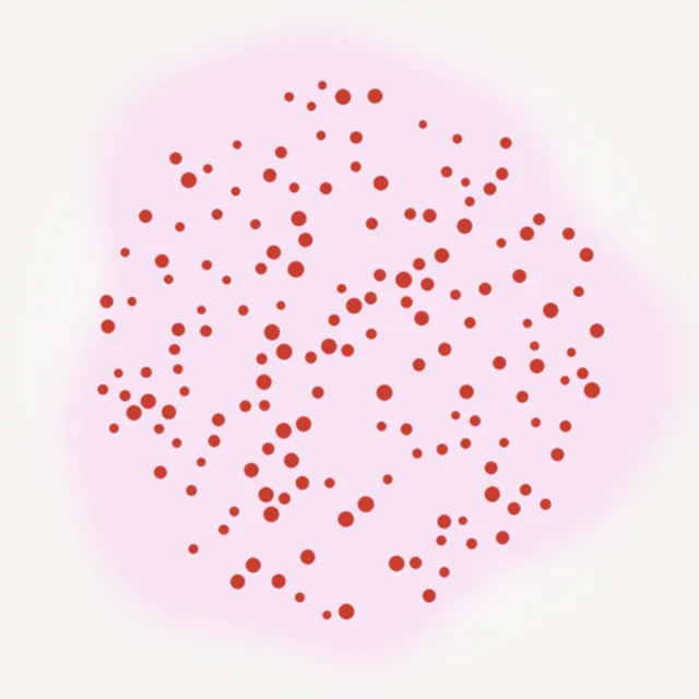 Schematic
Small red dots – vascular loops seen from above. Irregularly distributed in a melanocytic lesion they are suspicious; evenly distributed they are typical of psoriasis.
Typical of: Melanoma, Amelanotic melanoma, Psoriasis vulgaris
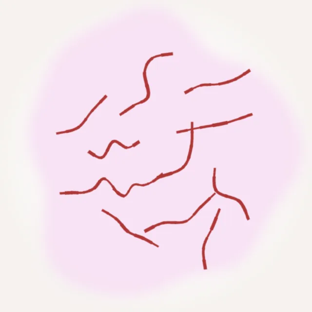 Schematic
Linear, irregularly winding vessels of varying calibre.
Typical of: Amelanotic melanoma, Melanoma, Basal cell carcinoma
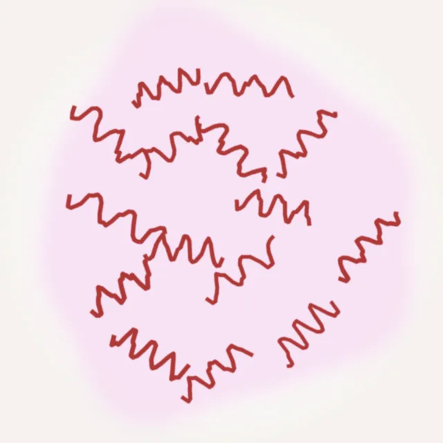 Schematic
Tightly spiralled vessels like a corkscrew.
Typical of: Amelanotic melanoma, Melanoma
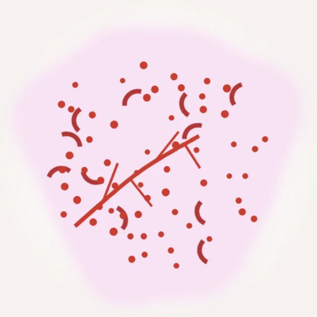 Schematic
Two or more different vessel types within one lesion, such as dots and serpentine lines.
Typical of: Amelanotic melanoma, Melanoma, Squamous cell carcinoma
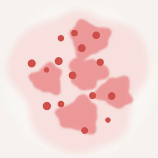 Schematic
Ill-defined milky pink to red areas or globules.
Typical of: Amelanotic melanoma, Melanoma
In the DermaFuchs dermoscopy course you find these structures on real images: tap the structure, name it, work out the diagnosis – free in the browser and as an app.
KLINIKFUCHS web app App Store More about DermaFuchsThis atlas is a learning resource and does not replace a medical examination. Whether a skin lesion is harmless can only be judged by a doctor examining it.