Green nail syndrome
- Synonyms
- chloronychia, pseudomonas nail infection
- Specialty
- Dermatology · Skin appendages
- Images
- Clinical 2 · Dermoscopy 3
- In the app
- 1 flashcard
- Last updated
- 10/2026 · Dr. Pascal Bafteh
Images (5)
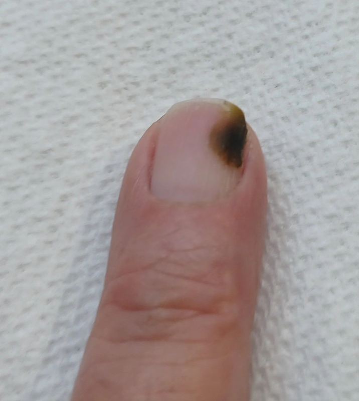
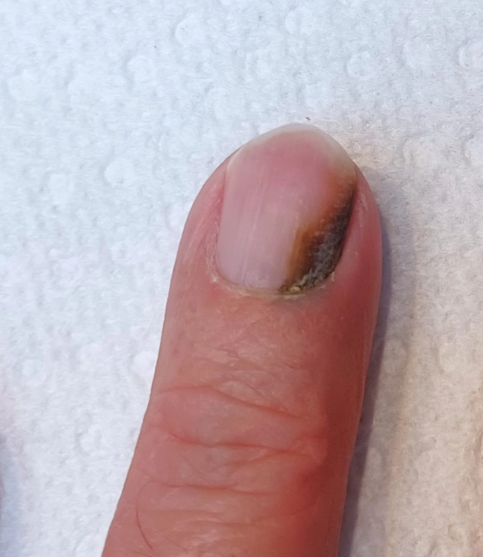
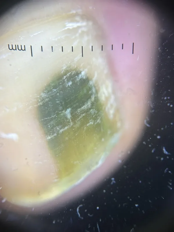 Dermoscopy
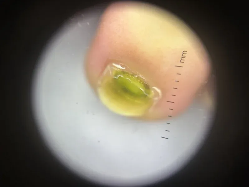 Dermoscopy
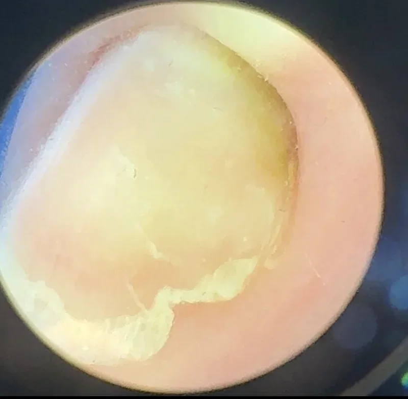 Dermoscopy
Definition
- Spot diagnosis: a green to blue-green-black discoloration of the nail plate, usually of a single nail with accompanying onycholysis or chronic paronychia, is a spot diagnosis. The colour sits within the nail plate and cannot be scraped off.
- Pathogen: The main causative agent is Pseudomonas aeruginosa with discoloration due to pyocyanin / pyoverdin.
- Predisposing factors: wet work, pre-existing onycholysis (portal of entry beneath the detached plate), chronic paronychia, onychomycosis, artificial nails, immunosuppression.
- Confirmation: under Wood's light the pyoverdin fluoresces greenish; if in doubt take a swab for culture (also for susceptibility testing). With accompanying onychodystrophy add a direct preparation and fungal culture, because onychomycosis and pseudomonal colonisation frequently coexist.
- Differential diagnoses: subungual haematoma (brownish-black, grows out with the nail), mould onychomycosis (Aspergillus, Scytalidium), melanonychia, exogenous staining by dyes.
Treatment principles
- The green discolouration is caused by Pseudomonas aeruginosa, usually in the space created by onycholysis; the most important measures are keeping the nails dry, avoiding wet work and trimming the detached part of the nail.
- Antiseptics or topical antibiotics are applied locally, and systemic therapy is required only exceptionally.
- Concomitant onychomycosis or nail psoriasis is treated as well.
Specific treatment regimens with drugs and doses are covered in the DermaFuchs app.
Keep learning in the app
Cross-references
More diseases of the skin appendages
Keep learning
Keep learning with flashcards, exam questions and spot diagnoses in the DermaFuchs app – free, in your browser or as an app.
Related pages: Hidrocystoma (eccrine and apocrine) · Lichen planopilaris · Milia (milium)
Note: This page is intended for medical education and does not replace diagnosis or treatment decisions in individual cases.