Halo naevus (Sutton naevus)
- Synonyms
- Sutton-Nävus, Leukoderma acquisitum centrifugum, Halonävus, halo naevus, halo nevus, Sutton naevus, mole with white ring
- Specialty
- Dermatology · Pigmentation disorders
- Images
- Clinical 2 · Histology 1
- In the app
- 1 flashcard
- Last updated
- 10/2026 · Dr. Pascal Bafteh
Contents
Images (3)
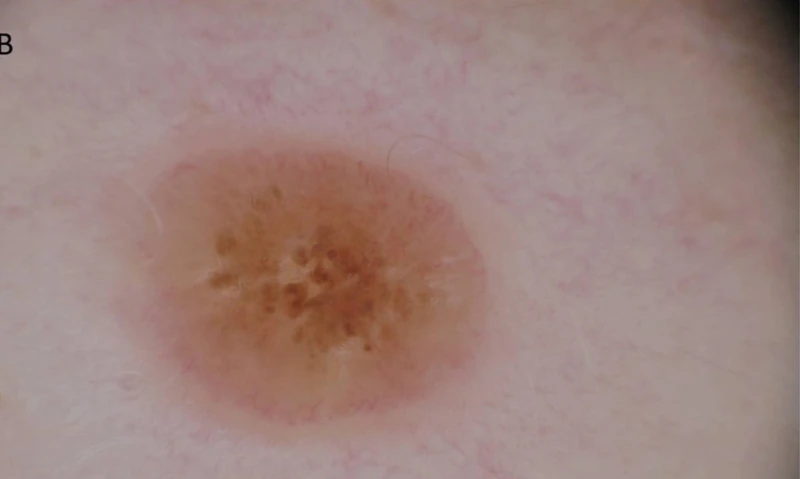
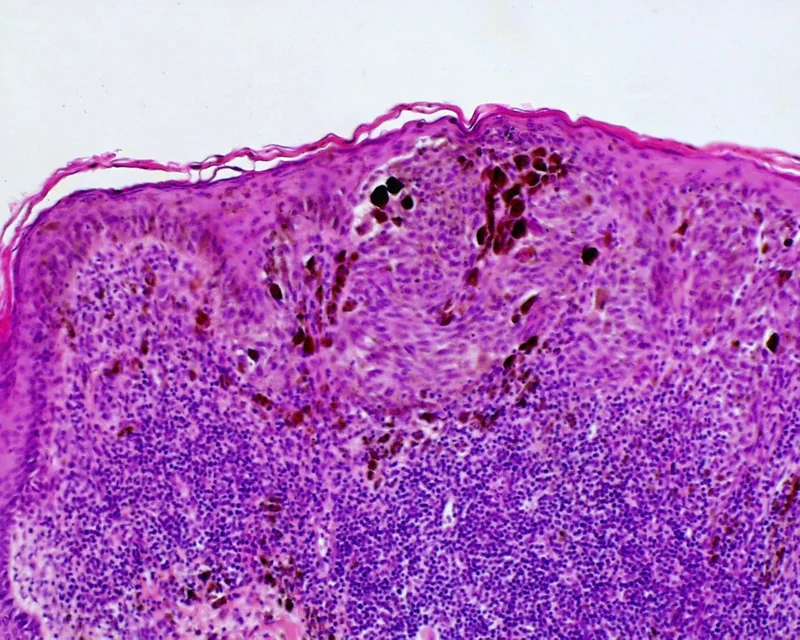 Histology
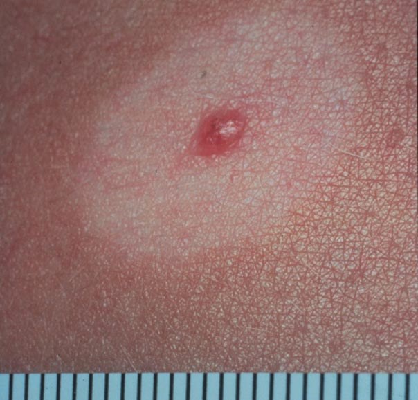
Definition
A halo naevus is a melanocytic naevus surrounded by a sharply demarcated depigmented ring. It is caused by a T-cell-mediated immune reaction against the naevus cells, which often leads to complete regression of the naevus.
Occurrence & epidemiology
Halo naevi are not uncommon (prevalence about 1% in white-skinned people) and occur mainly in healthy children and young adults, singly or multiply, mostly on the trunk. They are more common in people with vitiligo and other autoimmune diseases.
Clinical features
- Central, usually symmetrical brown naevus with a symmetrical, round to oval white halo usually 0.5–1 cm wide.
- Typical course in stages: the halo appears, the naevus fades and disappears, and the white area repigments partly or completely over years.
Diagnosis
- Clinical picture and dermoscopy (symmetrical central naevus, even depigmented halo).
- In adults, with an asymmetrical halo or an irregular central naevus, melanoma with regression is a differential diagnosis; total body examination (especially in adults), because halo naevi can rarely be triggered by a melanoma elsewhere.
Differential diagnoses
 Melanoma
 Vitiligo
 Psoriasis vulgaris (plaque psoriasis)
 Alopecia areata
 Amelanotic melanoma
 Dysplastic naevus (atypical naevus)
 Spitz naevus (spindle and epithelioid cell naevus)
Image credits for the thumbnails
- Melanoma: National Cancer Institute (unknown photographer), “Melanoma (4)”, Public Domain, via Wikimedia Commons · Public domain · Source
- Vitiligo: Klaus D. Peter, Gummersbach, Germany, “Vitiligo and Poliosis”, CC BY 3.0 de · CC BY 3.0 de · Source
- Psoriasis vulgaris (plaque psoriasis): MediaJet, “An Arm Covered With Plaque Type Psoriasis”, CC BY-SA 3.0, via Wikimedia Commons · CC BY-SA 3.0 · Source
- Alopecia areata: Abbassyma at English Wikipedia, “Allopecia areata”, Public Domain, via Wikimedia Commons · Public domain · Source
- Amelanotic melanoma: Dr. Thomas Brinkmeier, “Amelanotisches, malignes Melanom, ©wikiderm.de”, CC BY 4.0, via Wikimedia Commons · CC BY 4.0 · Source
- Dysplastic naevus (atypical naevus): 0x6adb015 (Wikimedia Commons), “DysplasticNevusSyndrome”, CC BY-SA 3.0, via Wikimedia Commons · CC BY-SA 3.0 · Source
- Spitz naevus (spindle and epithelioid cell naevus): The Armed Forces Institute of Pathology (AFIP), Public domain, via Wikimedia Commons · Public domain · Source
Treatment principles
- Halo naevus is benign and generally requires no treatment; the central naevus often regresses completely, while the depigmented zone may persist for a long time.
- Dermoscopic assessment of the central naevus and a total body skin examination are recommended, as a halo phenomenon can rarely occur around a melanoma or in association with a melanoma elsewhere.
- Excision is indicated only if the central component is atypical or the course is unusual, for example in older age.
Specific treatment regimens with drugs and doses are covered in the DermaFuchs app.
Keep learning in the app
References (selection)
Cross-references
Keep learning
Keep learning with flashcards, exam questions and spot diagnoses in the DermaFuchs app – free, in your browser or as an app.
Related pages: Melanoma · Vitiligo · Psoriasis vulgaris (plaque psoriasis)
Note: This page is intended for medical education and does not replace diagnosis or treatment decisions in individual cases.