Cutaneous amyloidoses
- Synonyms
- cutaneous amyloidosis, lichen amyloidosus, macular amyloidosis
- Specialty
- Dermatology · Metabolic skin disorders
- Images
- Clinical 1
- In the app
- 2 flashcards
- Last updated
- 10/2026 · Dr. Pascal Bafteh
Contents
Images (1)
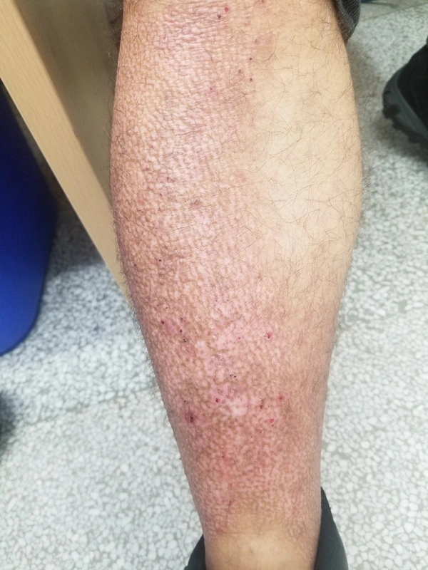
Definition
Definition: Deposition of misfolded proteins in the dermis, detected by Congo red staining with apple-green birefringence.
Amyloid Types:
- Amyloid K (keratinocytic)
- Amyloid L (immunoglobulin light chains)
- Amyloid A (acute-phase proteins)
Classification
Primary Cutaneous Amyloidoses
- Nodular form: Mostly amyloid L-type with risk of systemic involvement, produced by locally proliferating plasma cells; progression to systemic amyloidosis in about 10 % → investigation required.
Systemic Amyloidoses
Dermatological signs: Purpura, waxy papules, macroglossia, periorbital hematomas ("raccoon eyes").
Forms:
- AA amyloidosis: Reactive in chronic inflammatory diseases (e.g., IBD), often with renal involvement
Differential diagnoses
 Limited systemic sclerosis (CREST syndrome)
 Atopic dermatitis (atopic eczema)
 Prurigo nodularis
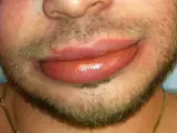 Angioedema
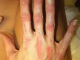 Dermatomyositis
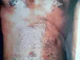 Darier's disease (keratosis follicularis)
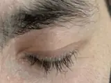 Lipoid proteinosis (Urbach–Wiethe disease)
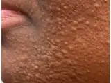 Lichen myxoedematosus (scleromyxoedema)
Image credits for the thumbnails
- Limited systemic sclerosis (CREST syndrome): James Heilman, MD, “CREST syndrome (calcinosis and sclerodactyly)”, CC BY-SA 4.0, via Wikimedia Commons · CC BY-SA 4.0 · Source
- Atopic dermatitis (atopic eczema): Eisfelder, “Atopic dermatitis child”, CC BY-SA 3.0, via Wikimedia Commons · CC BY-SA 3.0 · Source
- Prurigo nodularis: Chen R et al. (JAAD Case Rep 2026), Fig 1A, CC BY 4.0, via PubMed Central (PMC12723277) · CC BY 4.0 · Source
- Angioedema: James Heilman, MD, “AngioedemaFra”, CC BY-SA 3.0, via Wikimedia Commons · CC BY-SA 3.0 · Source
- Dermatomyositis: Mohammad2018, CC BY-SA 4.0 · CC BY-SA 4.0 · Source
- Darier's disease (keratosis follicularis): Mohammad2018, “Kerstosis follucularis”, CC BY-SA 4.0, via Wikimedia Commons · CC BY-SA 4.0 · Source
- Lipoid proteinosis (Urbach–Wiethe disease): Mobarak H, El Helou S (Cureus 2024), CC BY 4.0, via PubMed Central (PMC11463906) · CC BY 4.0 · Source
- Lichen myxoedematosus (scleromyxoedema): Kassalow B, Kazmi S, Shukri S et al. (Reports (MDPI) 2026), CC BY 4.0, via PubMed Central (PMC13214839) · CC BY 4.0 · Source
Treatment principles
- Treatment depends on the type; systemic amyloidosis must first be excluded, as it requires haematological and internal medicine management.
- In macular and lichen amyloidosis, controlling itch and breaking the scratch cycle are the priority, for example with potent topical glucocorticoids under occlusion and phototherapy.
- The nodular form can be excised or treated with ablative laser; recurrence is common in all forms.
Specific treatment regimens with drugs and doses are covered in the DermaFuchs app.
Keep learning in the app
References (selection)
Cross-references
Mentioned in
Keep learning: test yourself
One question from the DermaFuchs app on Cutaneous amyloidoses – tap an answer.
Question 1 of 1
Which type of amyloid is typically found in primary localized cutaneous amyloidosis (like macular or lichen amyloidosis)?
Answer
Correct: Amyloid K (keratinocytic). In primary localized cutaneous amyloidosis, the amyloid is derived from degenerated keratinocytes (Amyloid K).
More questions like these – with explanations and spaced repetition – in the app.
Note: This page is intended for medical education and does not replace diagnosis or treatment decisions in individual cases.