Lipoid proteinosis (Urbach–Wiethe disease)
- Synonyms
- Urbach-Wiethe-Syndrom, Hyalinosis cutis et mucosae, Lipoid proteinosis, Urbach-Wiethe disease
- Specialty
- Dermatology · Hereditary skin diseases
- Images
- Clinical 1 · Histology 1
- In the app
- 2 flashcards
- Last updated
- 10/2026 · Dr. Pascal Bafteh
Contents
Images (2)
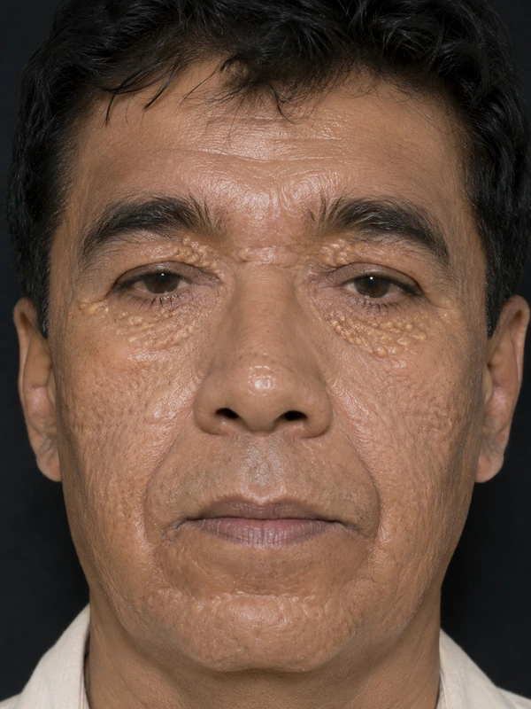 AI illustration
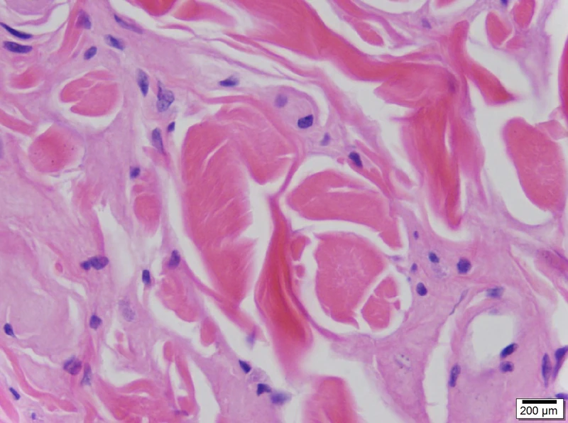 Histology
Definition
Lipoid proteinosis (Urbach–Wiethe disease, hyalinosis cutis et mucosae) is a very rare, autosomal recessive genodermatosis with progressive deposition of amorphous hyaline material in the skin, mucous membranes and internal organs. A typical early symptom is hoarseness starting in early childhood.
Occurrence & epidemiology
Just over 500 cases have been reported worldwide, with men and women equally affected; there is often a positive family history or parental consanguinity. The disease is more frequent in Namaqualand (South Africa) owing to a founder effect.
Aetiopathogenesis
It is caused by loss-of-function mutations in the gene for extracellular matrix protein 1 (ECM1, chromosome 1q21). ECM1 binds numerous matrix proteins; its loss disturbs the structure of skin and mucosa, with reduplicated basement membrane around vessels and at the dermal–epidermal junction.
Clinical features
- Hoarseness and a weak cry from infancy due to hyaline deposition in the laryngeal mucosa.
- Skin: first recurrent blisters on the face and limbs with pock-like scars; later yellowish, waxy papules and diffuse skin thickening, verrucous lesions on elbows, knees and knuckles.
- Moniliform blepharosis: bead-like yellowish papules along the eyelid margins, in about 50%.
- Mouth: thickened frenulum with limited tongue mobility, thickened lips.
- Central nervous system (50–75%): including epilepsy and behavioural changes, mainly due to calcification of the amygdala.
Histology
Homogeneous eosinophilic material in the dermis around vessels and adnexa; strongly PAS-positive, weakly positive or negative with Congo red.
Diagnosis
- Suspected from typical features (hoarseness since infancy, moniliform blepharosis, thickened frenulum).
- Biopsy: PAS-positive hyaline deposits.
- CT/MRI: bilateral bean-shaped calcifications of the amygdala are considered pathognomonic.
- Genetics: detection of biallelic ECM1 mutations confirms the diagnosis.
Differential diagnoses
Keep learning in the app
References (selection)
Cross-references
Note: This page is intended for medical education and does not replace diagnosis or treatment decisions in individual cases.