Porphyria cutanea tarda
Exam relevance: in 3 of more than 50 board exam reports · rank 45
- Synonyms
- PCT
- Specialty
- Dermatology · Metabolic skin disorders
- Images
- Clinical 1
- In the app
- 2 flashcards
- Last updated
- 10/2026 · Dr. Pascal Bafteh
Contents
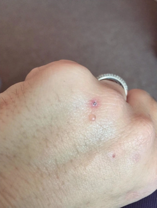
Images (1)
Definition
Porphyria cutanea tarda (PCT) is the most common porphyria. It results from reduced activity of hepatic uroporphyrinogen decarboxylase, so that porphyrins accumulate and make the skin photosensitive and mechanically fragile. An acquired (sporadic, type 1, about 75–80%) form is distinguished from a familial form (type 2, about 20–25%) with an autosomal dominant, heterozygous UROD mutation that becomes manifest only with additional triggers.
- Most common porphyria. Defect: Uroporphyrinogen decarboxylase. Triggers: Alcohol, estrogens, hepatitis C, iron.
- Clinical features: Blisters + erosions + milia on sun-exposed areas. Facial hypertrichosis, scleroderma-like changes.
- Diagnostics: Porphyrins in 24h urine (Uroporphyrin ↑↑). Wood's light: Coral-red fluorescence of urine.
Occurrence & epidemiology
Acquired PCT usually presents in the fourth or fifth decade, the familial form earlier. Precipitating factors are alcohol, oestrogens (contraceptives, hormone replacement), iron overload (e.g. haemochromatosis), hepatitis C (also B) and HIV infection, smoking and dialysis; several factors often act together.
Clinical features
- Increased fragility of sun-exposed skin, especially the back of the hands: blisters and erosions after minor trauma that leave crusts, scars and milia.
- Facial hypertrichosis, hyper- or hypopigmentation, sclerodermoid changes on the neck, face or chest; characteristically dark, reddish or tea-coloured urine.
- No acute photosensitive pain as in erythropoietic protoporphyria; no neurological attacks as in the acute hepatic porphyrias.
Histology
Subepidermal blister with PAS-positive deposits; the skin changes are histologically identical to those of variegate porphyria and hereditary coproporphyria – the diagnosis is confirmed by porphyrin analysis of blood, urine and faeces (diagnostic: raised faecal isocoproporphyrin).
Differential diagnoses
 Erythema ab igne
 Dermatomyositis
 Epidermolysis bullosa acquisita
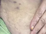 Erythema dyschromicum perstans (ashy dermatosis)
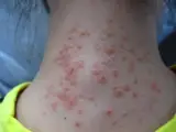 Hydroa vacciniforme
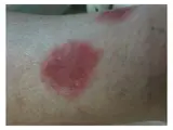 Pseudo-porphyria cutanea tarda
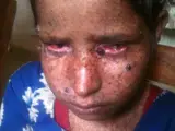 Xeroderma pigmentosum (XP)
- Porphyria cutanea tarda
Image credits for the thumbnails
- Erythema ab igne: James Heilman, MD, “Hotbottlerash”, CC BY-SA 3.0, via Wikimedia Commons · CC BY-SA 3.0 · Source
- Dermatomyositis: Mohammad2018, CC BY-SA 4.0 · CC BY-SA 4.0 · Source
- Epidermolysis bullosa acquisita: Szymański, Kowalewski, Pietrzyk et al. (2023), “Case Report: Biological treatment of epidermolysis bullosa acquisita: report on four cases and literature review”, CC BY 4.0, via Wikimedia Commons · CC BY 4.0 · Source
- Erythema dyschromicum perstans (ashy dermatosis): Golani S, Khan S, Saeed Z et al. (Cureus 2025), CC BY 4.0, via PubMed Central (PMC12168861) · CC BY 4.0 · Source
- Hydroa vacciniforme: Dr utsavsharma, “Hydroa-Vacciniforme-girl”, CC BY-SA 3.0, via Wikimedia Commons · CC BY-SA 3.0 · Source · cropped
- Pseudo-porphyria cutanea tarda: Katoulis AC, Ferra D, Toumbis E, Papadavid E, Kanelleas A, Panayiotides I, Rigopoulos D (Case Rep Dermatol Med 2013), “Pseudoporphyria associated with nonhemodialyzed renal insufficiency, successfully treated with oral N-acetylcysteine”, CC BY 3.0, via PubMed Central (PMC3639704) · CC BY 3.0 · Source
- Xeroderma pigmentosum (XP): Himynameislax, “Child suffering from Xeroderma Pigmentosa (Rukum Nepal)”, CC BY-SA 3.0, via Wikimedia Commons · CC BY-SA 3.0 · Source
Treatment principles
- The basis of treatment is avoiding triggers, particularly alcohol, oestrogens and iron intake, together with strict photoprotection that includes visible light.
- Venesection or antimalarials are used to reduce the porphyrin load.
- Associated conditions such as hepatitis C, HIV or haemochromatosis are actively sought and treated.
Specific treatment regimens with drugs and doses are covered in the DermaFuchs app.
Keep learning in the app
References (selection)
Cross-references
Mentioned in
Keep learning
Keep learning with flashcards, exam questions and spot diagnoses in the DermaFuchs app – free, in your browser or as an app.
Related pages: Erythema ab igne · Dermatomyositis · Epidermolysis bullosa acquisita
Note: This page is intended for medical education and does not replace diagnosis or treatment decisions in individual cases.