Hydroa vacciniforme
- Synonyms
- Hydroa vacciniformia Bazin, HV, hydroa vacciniforme-like lymphoproliferative disorder
- Specialty
- Dermatology · Environmental & photodermatoses
- Images
- Clinical 1
- In the app
- 1 flashcards
- Last updated
- 10/2026 · Dr. Pascal Bafteh
Contents
Images (1)
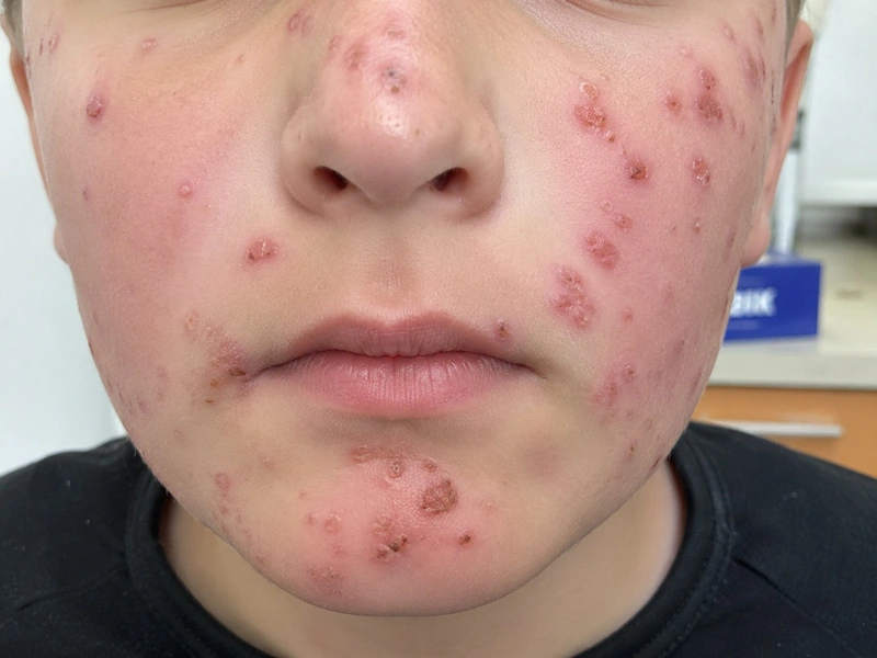 AI illustration
Definition
It is a very rare photodermatosis of childhood with recurrent vesicles on sun-exposed skin that heal with pox-like (varioliform) scars. It is now classified as a cutaneous form within the spectrum of Epstein-Barr virus (EBV)-associated T/NK-cell lymphoproliferative disorders.
Classification
- Classic form: benign, onset in childhood, papulovesicles on sun-exposed skin, mostly resolving by early adult life.
- Severe (systemic) form, termed "HV-like lymphoproliferative disorder" in the 2017 WHO classification: recurrent papulovesicular eruptions also on sun-protected skin, sometimes with facial oedema, fever, lymphadenopathy, oculomucosal lesions, gastrointestinal involvement and hepatosplenomegaly; progression to systemic T-cell or NK-cell lymphoma is possible. It is reported predominantly in children from Asia and Latin America.
Occurrence & epidemiology
It mostly affects children aged 3 to 15 years. It is diagnosed more often in girls; in boys it tends to start later and run a longer course.
Aetiopathogenesis
The trigger is ultraviolet light, most often long-wavelength UVA. The condition is frequently associated with chronic EBV infection; in the classic form, EBV-infected γδ T cells are increased.
Clinical features
- Sun-exposed sites: face, ears, hands, lower legs.
- About 30 minutes to 2 hours after sun exposure, burning, itching or stinging, followed by tense papules and vesicles, sometimes with facial swelling.
- Progression to umbilicated papules with black necrotic crusts on a red base; healing with pale depressed scars.
- First episode usually in spring, recurrences through the summer.
- Occasionally keratoconjunctivitis, photophobia and photo-onycholysis.
Histology
Spongiosis, vesiculation and epidermal necrosis overlying a dense dermal infiltrate; scarring in older lesions. EBV can be detected in the dermal infiltrate by EBER in situ hybridisation.
Diagnosis
- Clinical suspicion from the typical history and distribution; skin biopsy with EBER in situ hybridisation.
- Determine the EBV status.
- Look for signs of the severe form: involvement of sun-protected skin, fever, lymphadenopathy, hepatosplenomegaly.
Differential diagnoses
Keep learning in the app
References (selection)
- DermNet: Hydroa vacciniforme
- DermNet: Hydroa vacciniforme pathology
- PubMed: Hydroa Vacciniforme and Hydroa Vacciniforme-Like Lymphoproliferative Disorder: A Spectrum of Disease Phenotypes Associated with Ultraviolet Irradiation and Chronic Epstein-Barr Virus Infection (PMID 33297336)
- PubMed: Hydroa vacciniforme: a distinctive form of Epstein-Barr virus-associated T-cell lymphoproliferative disorders (PMID 30998212)
Cross-references
Note: This page is intended for medical education and does not replace diagnosis or treatment decisions in individual cases.