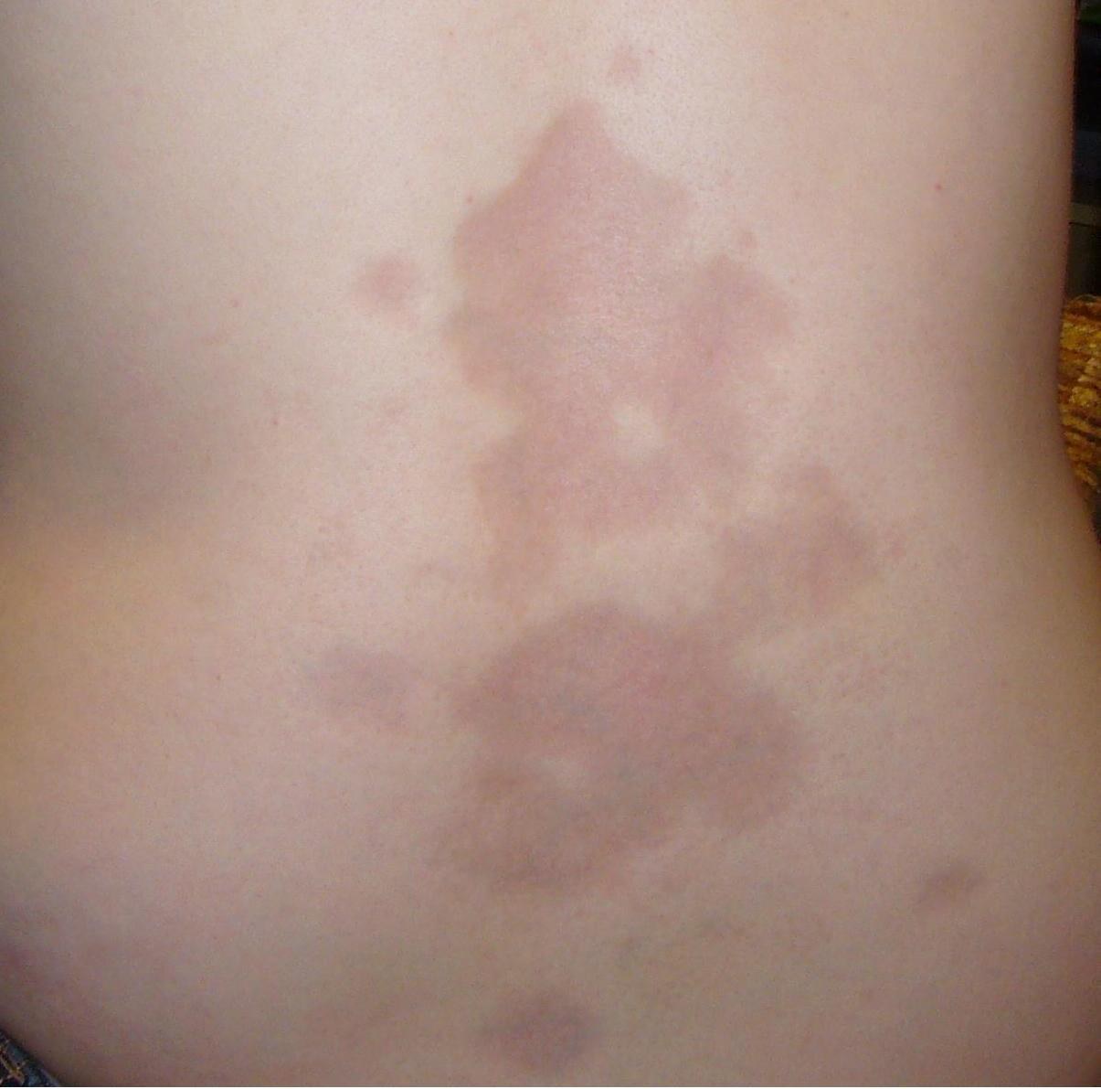
Morphea
Overview
- No systemic involvement. Variants: Plaque morphea, linear morphea (en coup de sabre), generalized morphea.
- Morphea Dermoscopy: Fibrotic beams (white rays). Lilac ring = active border.
- Circumscribed Scleroderma en coup de sabre: Linear morphea in the forehead area. Can affect deeper structures (Parry-Romberg).
- Nailfold Capillaroscopy: In SSc megacapillaries + avascular areas. Earliest clinical marker.
- 20 MHz high-frequency ultrasound: The standard technique for measuring dermal thickness and echogenicity in morphea and systemic sclerosis — active lesions show a thickened, hypoechoic dermis, burnt-out atrophic lesions a thin, hyperechoic dermis, which makes the disease course measurable. The same 20 MHz technique is used preoperatively in melanoma to estimate tumour thickness for planning the excision margin; the sonographic thickness usually exceeds the histological Breslow thickness because the peritumoural inflammatory infiltrate is measured as well.
Differential diagnoses
- Limited systemic sclerosis (CREST syndrome)
- Scleredema Buschke
- Dermatomyositis
- Lichen sclerosus
- Sarcoidosis
- Psoriasis vulgaris (plaque psoriasis)
- Atypical fibroxanthoma
- Cutaneous angiosarcoma
Practise Morphea in the app
Flashcards with spaced repetition, exam questions and spot-the-diagnosis on this topic – in DermaFuchs, free of charge.
In the DermaFuchs app: 6 flashcards · 6 clinical images
Open in browser Download on the App StoreSources (selection)
More connective tissue diseases
Note: This page is intended for medical education and does not replace diagnosis or treatment decisions in individual cases. Treatment and follow-up content is available in the app.