Scabies
Definition & Pathogen
- Pathogen: Sarcoptes scabiei var. hominis — obligate human pathogenic mite that burrows into the stratum corneum and creates tunnels.
- Biology: Female lives 4–6 weeks, lays 2–3 eggs daily in the stratum corneum; larvae hatch after 3–4 days.
- Transmission: close skin contact (≥ 5–10 min), to a lesser extent via textiles (bed linen, clothing).
Clinical Presentation & Predilection Sites
- Cardinal symptom: intense pruritus, worse at night.
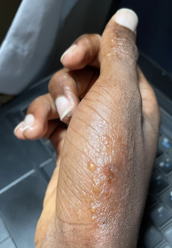
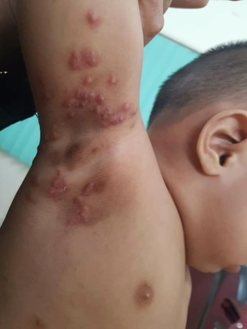
- Lesions: comma-shaped mite burrows, papules, vesicles, excoriations, secondary eczematous changes.
- Predilection: interdigital folds of the hands, flexor wrists, axilla, areolae, periumbilical, penis/scrotum, medial malleoli — usually sparing the face (except in infants).
Diagnostics
- Dermatoscopy: reveals the delta sign + ink sign.
- Light examination: in Wood's light, burrows appear white.
- Microscopy: skin scraping/adhesive tape sample → detection of scybala, mites & eggs.
General & Environmental Measures
- Laundry: wash clothes/bed linen at > 50 °C or 4 days in plastic bag.
- Mattresses: infestation affects only the cover.
- Contact persons: simultaneous treatment of all household members and close contacts (even if asymptomatic).
Special form: Crusted Scabies
- Crusted scabies (Norwegian scabies) = extreme variant in immunosuppressed individuals (HIV, hematologic comorbidity, glucocorticoid therapy).
- Highly contagious due to millions of mites; widespread hyperkeratotic plaques, pruritus often reduced.
First step: isolation
Before any work-up or treatment comes immediate contact isolation — with a mite burden of up to several million, brief skin contact as well as contaminated linen and surfaces suffice for transmission. This is why crusted scabies is the classic trigger of outbreaks in care homes and hospitals.
- Single room, gowns and gloves for every contact, rigorous processing of linen and surfaces; contacts — fellow residents, nursing staff, relatives — are treated simultaneously (see section 9).
Confirming the diagnosis: direct preparation from the scales
In crusted scabies the laborious search for burrows is unnecessary: in a microscopic direct preparation of detached horny scales a single scale already contains numerous mites, eggs and scybala.
- Practical: remove scales with a scalpel or curette, place them in potassium hydroxide or immersion oil on a slide, apply a coverslip and examine; dermoscopy and adhesive tape sampling are complementary.
- Look for the cause: HIV test, haematological work-up, drug history (glucocorticoids, immunosuppressants), nursing and nutritional status.
Differential diagnoses
- Impetigo
- Dyshidrotic hand eczema (pompholyx)
- Urticaria papulosa
- Allergic contact dermatitis
- Scabies
- Atopic dermatitis (atopic eczema)
- Cimicosis (bed bug bites)
- Demodicosis
Practise Scabies in the app
Flashcards with spaced repetition, exam questions and spot-the-diagnosis on this topic – in DermaFuchs, free of charge.
In the DermaFuchs app: 8 flashcards · 11 clinical images
Open in browser Download on the App StoreSources (selection)
- Chandler et al. 2019 — Scabies
- AWMF S1 Skabies 2023
- Leung et al. 2019 — Scabies a neglected global disease
More parasitic skin diseases
- Demodicosis
- Filariasis
- Cutaneous leishmaniasis
- Cutaneous larva migrans
- Pediculosis capitis
- Caterpillar dermatitis caused by oak processionary moth (Thaumetopoea processionea)
- Crusted (Norwegian) scabies
- Tungiasis
Note: This page is intended for medical education and does not replace diagnosis or treatment decisions in individual cases. Treatment and follow-up content is available in the app.