Cholesterol embolism (atheroembolism)
- Synonyms
- Cholesterinkristallembolie, Atheroembolie, Blue-toe-Syndrom, Purple-toe-Syndrom, cholesterol crystal embolism, atheroembolism, blue toe syndrome
- Specialty
- Dermatology · Vascular skin disorders
- Images
- Clinical 1 · Histology 1
- In the app
- 1 flashcards
- Last updated
- 10/2026 · Dr. Pascal Bafteh
Contents
Images (2)
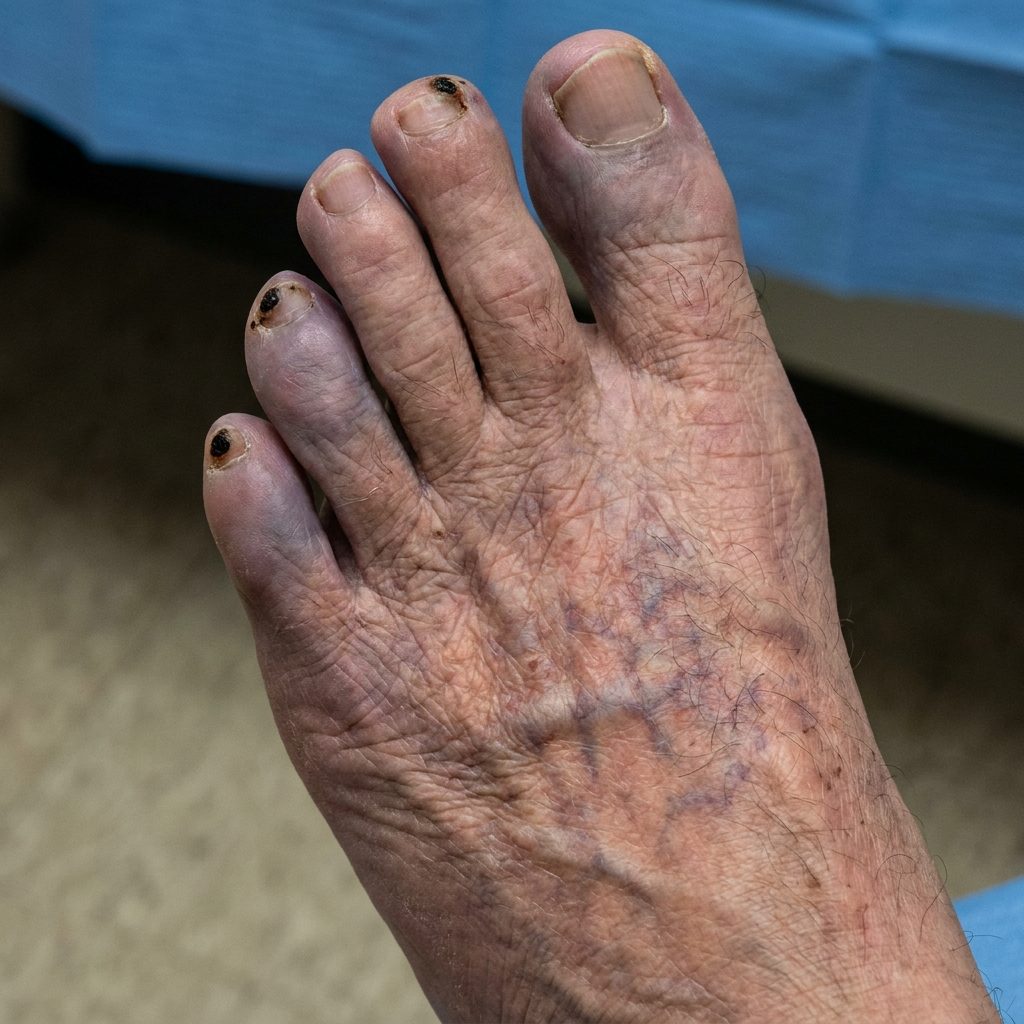 AI illustration
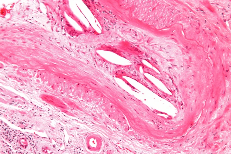 Histology
Definition
In cholesterol embolism, cholesterol crystals break off from ulcerated atherosclerotic plaques in large arteries (e.g. the aorta, iliac and femoral arteries) and occlude small arteries of the skin and internal organs. The result is impaired blood flow up to tissue death. Cholesterol embolism is the most common cause of blue toe syndrome.
Aetiopathogenesis
People with atherosclerosis are affected. Embolisation can occur spontaneously but most often follows hours to days after a vascular procedure that disrupts the plaque surface (after up to 1% of such procedures), e.g. angiography, angioplasty, intra-aortic balloon pump or cardiopulmonary resuscitation. It has rarely been reported after thrombolysis or with drugs that inhibit blood clotting.
Clinical features
- The skin is involved in over one third of patients: livedo reticularis or racemosa, gangrene, acrocyanosis ("blue toes"), ulceration, painful nodules and purpura. In a series of 223 patients, those with skin involvement had livedo in 49%, gangrene in 35% and acrocyanosis in 28%.
- Affected toes are blue-purple, cold and acutely painful, even at rest; foot pulses may be palpable because small arteries and arterioles are occluded.
- Organ involvement: acute kidney failure (25–50%), retinal ischaemia, pancreatitis, intestinal infarction; plus non-specific fever, myalgia and weight loss.
Histology
Small arteries contain cholesterol clefts – empty spaces, because the crystals are dissolved during tissue fixation – often together with thrombi, macrophages, giant cells and eosinophils.
Diagnosis
- Clinical suspicion in known atherosclerosis with typical skin signs, worsening kidney function or abdominal symptoms, particularly after a vascular procedure.
- The diagnosis is confirmed histologically by biopsy of affected skin or other tissue.
- Laboratory: eosinophilia in up to 80%, sometimes raised white cell count and ESR.
- Fundoscopy: Hollenhorst plaques.
Differential diagnoses
- Arterial (ischaemic) ulcer
- Chronic venous insufficiency
- Necrotizing (leukocytoclastic) vasculitis
- Livedo reticularis
- Warfarin-induced skin necrosis (coumarin necrosis)
Keep learning in the app
References (selection)
Cross-references
Note: This page is intended for medical education and does not replace diagnosis or treatment decisions in individual cases.