Keloid
- Synonyms
- Wulstnarbe, Narbenkeloid, hypertrophe Narbe, Keloidnarbe, keloid scar, hypertrophic scar
- Specialty
- Dermatology · Environmental & photodermatoses
- Images
- Clinical 9 · Histology 3
- In the app
- 4 flashcards
- Last updated
- 10/2026 · Dr. Pascal Bafteh
Contents
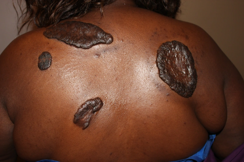
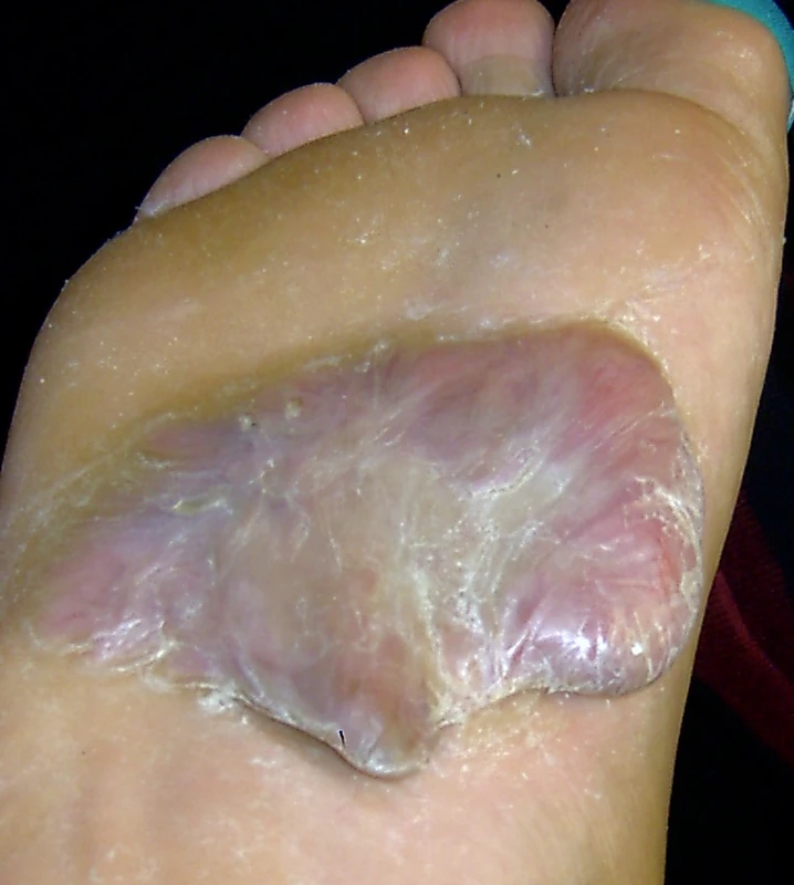
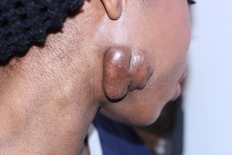
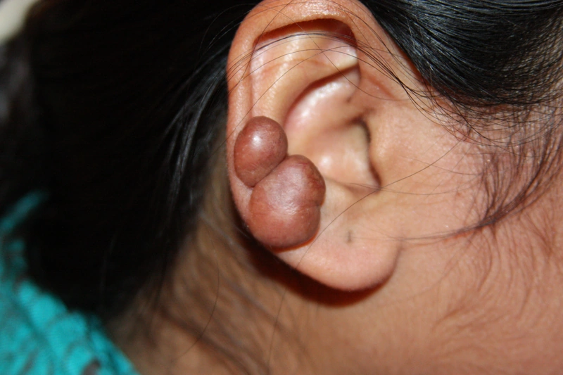
Images (12)
 Histology
 Histology
 Histology
Definition
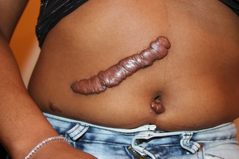
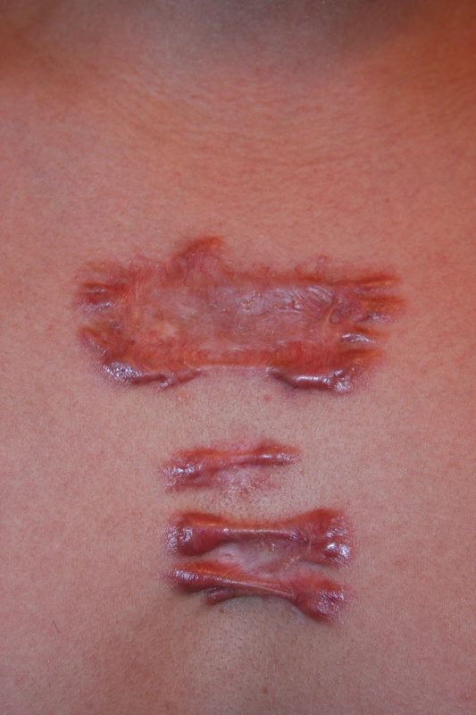
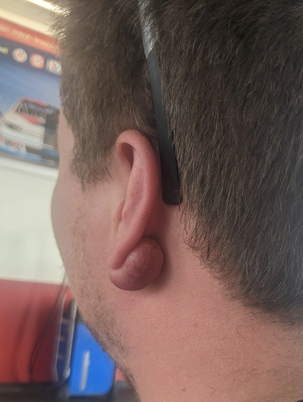
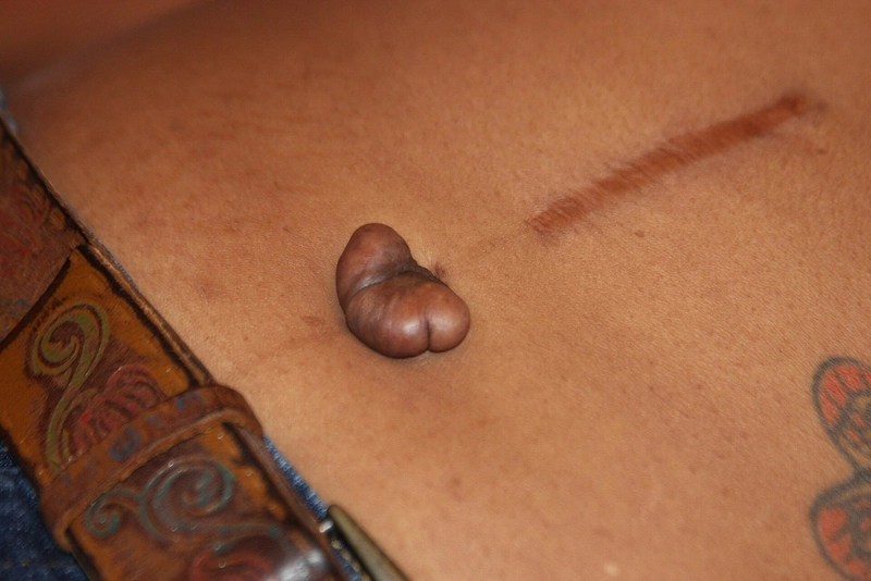
A keloid is an excessive benign connective tissue overgrowth that extends beyond the boundaries of the original wound after skin injury. It differs from a hypertrophic scar, which remains confined to the wound, usually develops within weeks of the injury and is more likely to regress.
Occurrence & epidemiology
Keloids are more frequent in Fitzpatrick skin types III to VI; a genetic association (certain HLA haplotypes, blood group A) has been described. Even minor injuries can be triggers, e.g. cuts, burns, insect bites, acne or infections such as herpes zoster; rarely they arise spontaneously, sometimes only months to years after the injury.
Clinical features
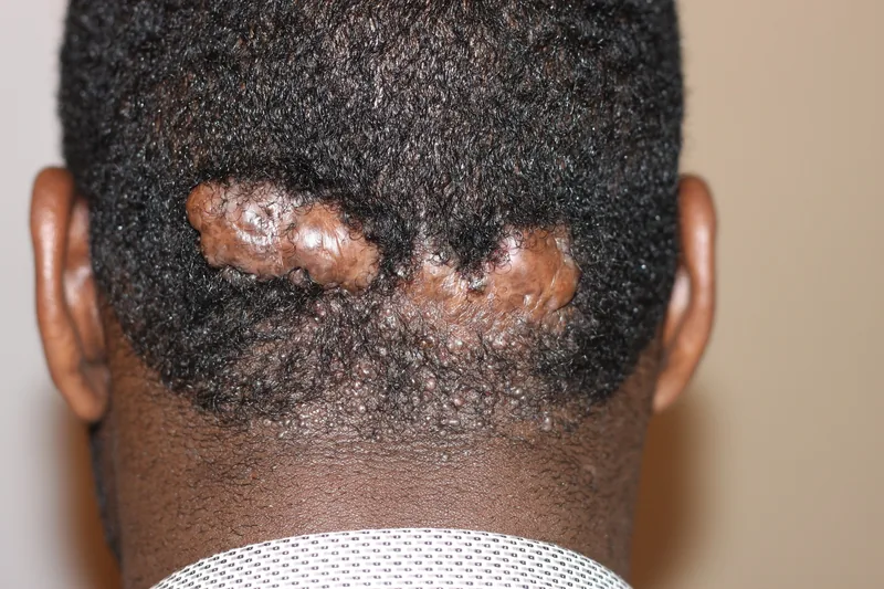
- Firm, smooth, raised, purplish-red ridges (more pigmented on dark skin) that grow beyond the wound area.
- May be itchy or uncomfortable; keloids tend to persist.
- Sites of predilection (areas of high skin tension): upper chest, shoulders, earlobes, neck, upper arms and upper back.
Histology
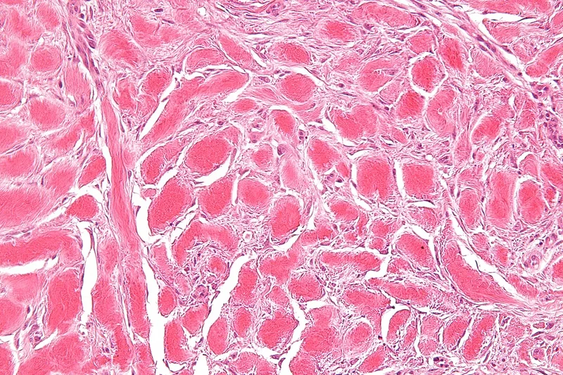
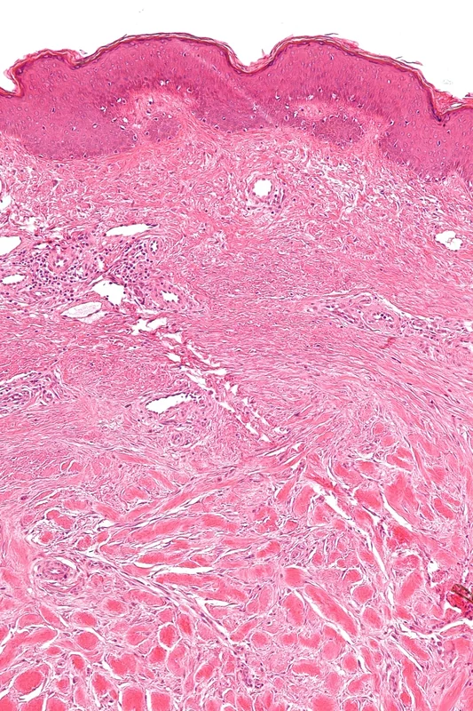
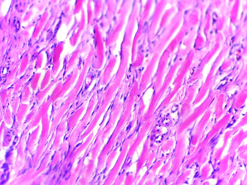
Whorls and nodules of particularly thick, homogeneous collagen bundles scattered irregularly throughout the dermis ("keloidal collagen") – absent, however, in up to half of keloids; increased fibroblasts. Hypertrophic scars show increased fibroblasts and a higher density of collagen fibres.
Diagnosis
- Clinical diagnosis based on history, extension beyond the wound and course.
- Biopsy if the diagnosis is uncertain, among other things to rule out dermatofibrosarcoma protuberans, squamous cell carcinoma, pseudolymphoma and other tumours.
Treatment principles
- Keloids are highly prone to recurrence; excision alone is therefore not appropriate.
- First-line treatment is intralesional glucocorticoids, often combined with cryotherapy; silicone sheeting and compression are used as adjuncts.
- If excision is performed, it is always followed by adjuvant treatment, such as intralesional glucocorticoids or radiotherapy; laser procedures can improve redness and volume.
- In patients with a known tendency to keloids, elective procedures and piercings should be weighed carefully.
Specific treatment regimens with drugs and doses are covered in the DermaFuchs app.
Keep learning in the app
References (selection)
Cross-references
Keep learning
Keep learning with flashcards, exam questions and spot diagnoses in the DermaFuchs app – free, in your browser or as an app.
Related pages: Livedo racemosa · Miliaria (heat rash, prickly heat) · Miliaria Profunda
Note: This page is intended for medical education and does not replace diagnosis or treatment decisions in individual cases.