Lymphoedema
- Synonyms
- Lymphoedem, Lymphstauung, primäres Lymphödem, sekundäres Lymphödem, Stemmer-Zeichen, Elephantiasis nostras verrucosa, lymphedema, primary lymphoedema, secondary lymphoedema, Stemmer sign
- Specialty
- Dermatology · Vascular skin disorders
- Images
- Clinical 1
- In the app
- 8 flashcards
- Last updated
- 10/2026 · Dr. Pascal Bafteh
Contents
Images (1)
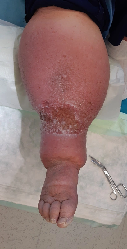
Definition
Lymphoedema is chronic swelling caused by impaired lymph flow: protein- and water-rich tissue fluid is not adequately carried away. It may be localised or diffuse, e.g. involving one or more limbs.
Classification
- Primary lymphoedema: aplasia or hypoplasia of the lymphatic vessels, often inherited; by age at onset congenital (before age 2, e.g. autosomal dominant Milroy disease with VEGFR3 mutations), lymphoedema praecox (age 2–35, typically in females at menarche or pregnancy) and lymphoedema tarda (after age 35).
- Secondary lymphoedema (far more common): disruption of lymphatic vessels, e.g. after lymph node dissection or radiogenic damage in cancer, in the tropics mainly due to lymphatic filariasis; also with chronic venous insufficiency, trauma, cellulitis and tumour obstruction.
Clinical features
- Sensation of tightness and heaviness; usually more pronounced in the legs than in the arms.
- Stage 1: pitting oedema that often subsides overnight.
- Stage 2: non-pitting oedema, early fibrosis due to chronic inflammation.
- Stage 3: brawny, irreversible oedema due to soft-tissue fibrosis, thickened skin with a peau d'orange appearance.
- With progression deep skin folds and warty papules on the ankles, feet and toes (elephantiasis nostras verrucosa); extreme forms (elephantiasis) mainly in filariasis.
- Complications: cellulitis and other bacterial infections, dermatitis, rarely lymphangiosarcoma (Stewart-Treves syndrome).
Diagnosis
- Mainly a clinical diagnosis based on history (including family history) and examination.
- Stemmer sign: positive if the skin over the dorsum of the foot or hand cannot be pinched into a fold. In a lymphoscintigraphy-controlled study, sensitivity was 92% but specificity only 57% (false positives e.g. in obesity).
- Lymphoscintigraphy when the diagnosis or cause is unclear; CT/MRI show sites of obstruction; ultrasound to exclude e.g. deep vein thrombosis; search for a tumour if no cause is found.
- Distinction from lipoedema (fat accumulation in both thighs and lower legs).
Differential diagnoses
- Great saphenous vein varicosity
- Purpura jaune d’ocre
- CRPS / Sudeck atrophy
Keep learning in the app
References (selection)
Cross-references
Note: This page is intended for medical education and does not replace diagnosis or treatment decisions in individual cases.