Lichen aureus
- Synonyms
- Martin 1958, pigmented purpuric dermatosis
- Specialty
- Dermatology · Vascular skin disorders
- Images
- Clinical 2
- In the app
- 1 flashcard
- Last updated
- 10/2026 · Dr. Pascal Bafteh
Contents
Images (2)
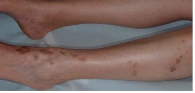
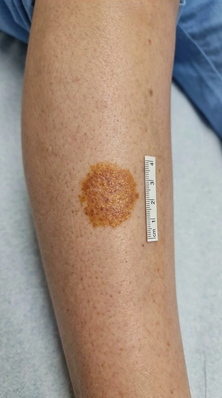 AI illustration
Definition
Definition & Classification
- Pigmented purpuric dermatoses (PPD; purpura pigmentosa progressiva, "capillaritides") = group of chronic, benign lymphocytic capillaritides with erythrocyte extravasation and hemosiderin deposition — not a true vasculitis (no fibrinoid vessel-wall necrosis).
- Hallmark: non-blanchable (diascopy negative) petechial "cayenne-pepper" dots + golden-brown/orange hemosiderin macules, preferentially on the lower legs/pretibial, usually symmetrical, course over months to years.
- Schamberg disease mostly affects older men, lichen aureus preferentially children and young adults; often asymptomatic or mildly itchy.
Aetiopathogenesis
Pathophysiology — WHY the golden-brown color?
Cell-mediated (predominantly CD4⁺ T-cell) inflammation around the postcapillary venules/capillaries of the papillary dermis → endothelial swelling, increased vascular permeability and fragility → extravasation of erythrocytes into the corium (fresh lesion = bright red) → phagocytosis by macrophages → intracellular hemosiderin (Fe³⁺) in siderophages → golden-brown "aureus" hue (Perls/Prussian blue positive). There is no fibrinoid necrosis and leukocytoclasia — this is exactly what histologically separates PPD from leukocytoclastic vasculitis.
Clinical features
Clinical Variants (Eponyms)
- Schamberg disease (purpura pigmentosa progressiva, Schamberg 1901) — most common form: cayenne-pepper petechiae on a brownish background, lower legs.
- Purpura annularis telangiectodes (Majocchi 1896) — annular/ring-shaped lesions with telangiectasias at the border and central clearing, occasionally mild atrophy.
- Lichenoid purpura (Gougerot-Blum 1925) — small lichenoid papules, purpuric, confluent.
- Eczematid-like purpura (Doucas-Kapetanakis 1953) — with scaling and marked pruritus.
- Lichen aureus (lichen purpuricus, Martin 1958) — usually solitary, golden-yellow to rust-brown group of lichenoid papules, often on the leg, frequently overlying a varicose vein, particularly persistent.
Diagnosis
Diagnostics & Histology
- Diascopy (glass-spatula pressure): purpura remains visible (non-blanchable) — distinction from erythema/telangiectasia.
- Histopathology: superficial perivascular lymphocytic infiltration, erythrocyte extravasation, hemosiderin detection (Perls/Prussian blue +); no leukocytoclasia/fibrinoid necrosis.
- Dermoscopy: reddish-brown dots/globules on a coppery/orange-brown background.
Differential diagnoses

- Schamberg's disease (progressive pigmented purpura)
- Purpura annularis telangiectodes (Majocchi disease)
- Pigmented purpuric lichenoid dermatosis of Gougerot-Blum
Image credits for the thumbnails
- Mycosis fungoides: Wikindrea, “Mycosis Fungoides Before Sun and Cartamo”, CC BY-SA 4.0, via Wikimedia Commons · CC BY-SA 4.0 · Source
- Leukocytoclastic vasculitis: James Heilman, MD, “LV22010 (leukocytoclastic vasculitis)”, CC BY-SA 3.0, via Wikimedia Commons · CC BY-SA 3.0 · Source
- Kaposi's sarcoma: unknown author, “Kaposi's Sarcoma”, Public Domain, via Wikimedia Commons · Public Domain · Source
Treatment principles
- Lichen aureus is benign and runs a chronic course, sometimes with spontaneous regression; patient education is the priority.
- Possible triggers such as drugs are avoided, and compression therapy is used when there is a venous component.
- For itch, short-term moderate-potency topical glucocorticoids may be considered, and phototherapy for extensive or persistent forms.
Specific treatment regimens with drugs and doses are covered in the DermaFuchs app.
Keep learning in the app
References (selection)
- Plewig G, French L, Ruzicka T, Kaufmann R, Hertl M (Hrsg). Braun-Falcos Dermatologie, Venerologie und Allergologie. 7. Aufl. Berlin: Springer; 2018. S. 1252–1254.
- Pigmented Purpuric Dermatoses: A Complete Narrative Review
- Tolaymat L, Hall MR. Pigmented Purpuric Dermatosis. In: StatPearls [Internet]. Treasure Island (FL): StatPearls Publishing; 2023.
- Pigmented purpuric dermatosis or mycosis fungoides: A diagnostic dilemma
Cross-references
Keep learning
Keep learning with flashcards, exam questions and spot diagnoses in the DermaFuchs app – free, in your browser or as an app.
Related pages: Mycosis fungoides · Leukocytoclastic vasculitis · Kaposi's sarcoma
Note: This page is intended for medical education and does not replace diagnosis or treatment decisions in individual cases.